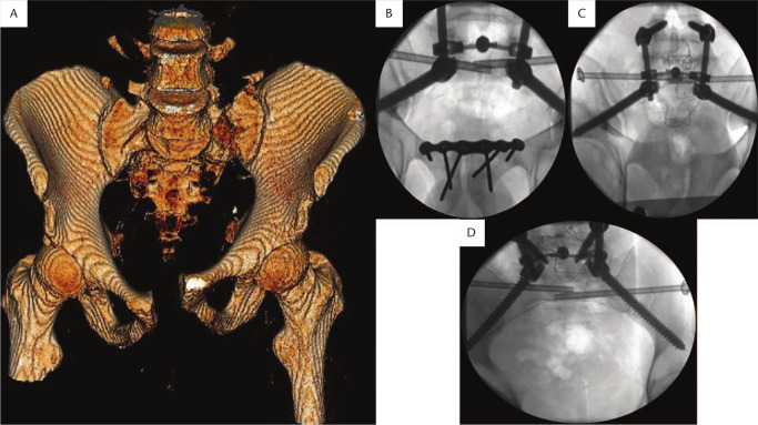
Fig. 8.
(A) Three-dimensional pelvic model showing bilateral sacral fractures and pubis diastasis anteriorly. (B) Anteroposterior, (C) outlet, and (D) inlet fluoroscopic views showing stabilization of the fracture with spinopelvic fixation and sacroiliac screws to S1 body (triangular configuration). Pubis symphysis was stabilized with plating anteriorly.

