
Figure 3. Patched traffics from the apical to the basal side of the wing disc through Multivesicular Bodies.
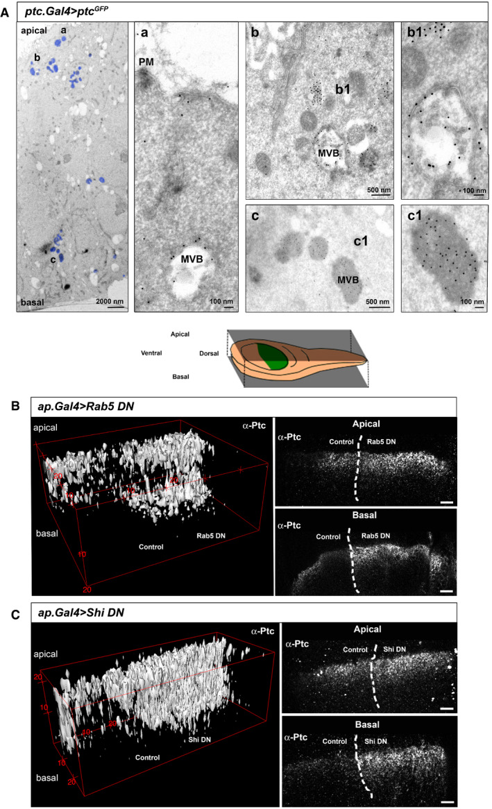
- Electron microscopy imaging of wing discs expressing the UAS‐Ptc‐GFP construct for 24 h in receiving cells using Ptc‐Gal4; TubGal80ts. Left panel shows areas with anti‐GFP immuno‐labelling (marked in blue) throughout the apico/basal axis of the wing disc epithelium (a) anti‐GFP gold labelling corresponding to Ptc is located on the apical membrane and early endosomes towards MVB formation. (b) Ptc immunogold labelling is present in multivesicular bodies (MVBs) subapically (top right panels). b1) Note that subapical MVBs appear as less dense with greater Ptc labelling on the MVB outer membranes. (c) Ptc label is also present in MVBs close to the basal membrane (lower right panels), (c1) Note that basolateral Ptc MVBs are significantly denser and richer in Ptc positive intraluminal vesicles (ILV).
- Endogenous Ptc subcellular localization after endocytosis inhibition by the dorsal expression of a dominant negative form of Rab5. The top panel is a schematic representation of the localized expression. The left panel is a digital 3D reconstruction and the right panel shows an apical and basal confocal sections. Note that accumulation of Ptc occurs at both the apical and basal sides of the dorsal part of the disc, compared to the wild‐type control on the ventral side.
- Digital 3D reconstruction (left) and apical and basal confocal sections (right) of endogenous Ptc localization after endocytosis inhibition by the dorsal expression of a dominant negative form of shibire (Shi DN). Note the same apical and basal accumulation as after expression of Rab5DN, and comparing to the ventral wild‐type half.