-
A
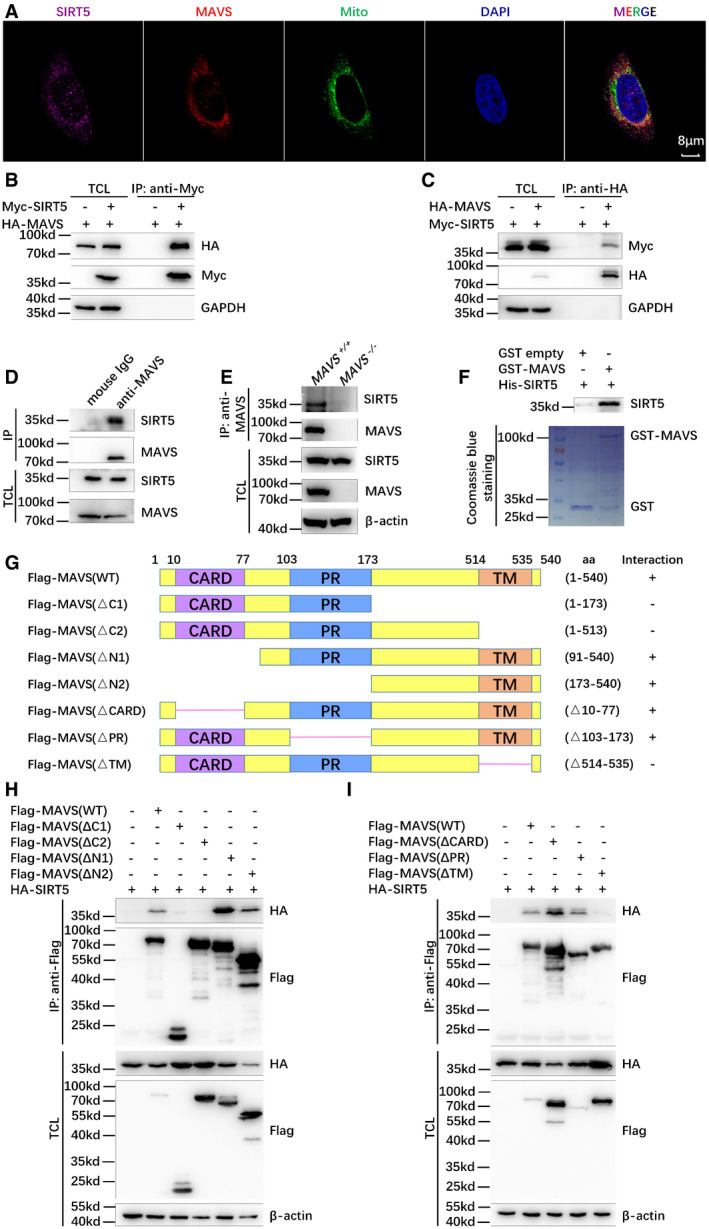
Confocal microscopy image of endogenous SIRT5 co‐localized with endogenous MAVS in H1299 cells detected by immunofluorescence staining using anti‐SIRT5 and anti‐MAVS antibodies. Mito, MitoTracker; Scale bar = 8 μm.
-
B, C
Co‐immunoprecipitation of Myc‐SIRT5 with HA‐MAVS and vice versa. HEK293T cells were co‐transfected with indicated plasmids for 24 h. Anti‐Myc (B) or anti‐HA antibody‐conjugated agarose beads (C) were used for immunoprecipitation, and the interaction was detected by immunoblotting with the indicated antibodies.
-
D
Endogenous interaction between MAVS and SIRT5. Anti‐MAVS antibody was used for immunoprecipitation, and normal mouse IgG was used as a control.
-
E
Endogenous interaction between MAVS and SIRT5 in the wild‐type (WT) (MAVS
+/+) or SIRT5‐deficient (MAVS
−/−) H1299 cells. Anti‐MAVS antibody was used for immunoprecipitation, and the interaction was detected by immunoblotting with anti‐SIRT5 antibody.
-
F
GST pull‐down assay for GST‐tagged MAVS and His‐tagged SIRT5. GST‐tagged MAVS and His‐tagged SIRT5 were expressed in Escherichia coli (BL21), respectively. The association of GST‐MAVS with His‐SIRT5 was detected by immunoblotting with anti‐SIRT5 antibody. GST and GST‐MAVS proteins were stained with Coomassie blue.
-
G
Schematic of MAVS domains interacted with SIRT5. The interaction is indicated by plus (+) sign.
-
H, I
Co‐immunoprecipitation analysis of HA‐SIRT5 with Flag‐MAVS‐truncated mutants. HEK293T cells were co‐transfected with the indicated plasmids. Anti‐Flag antibody‐conjugated agarose beads were used for immunoprecipitation, and the interaction was analyzed by immunoblotting with the indicated antibodies. Flag‐MAVS fragments (WT: full length; ΔC1, 1–173 aa; ΔC2, 1–513 aa; ΔN1, 91–540 aa; ΔN2, 173–540 aa; ΔCARD, Δ10–77 aa; ΔPR, Δ103–173 aa; ΔTM, Δ514–535 aa).
Data information: IP, immunoprecipitation; TCL, total cell lysates.

