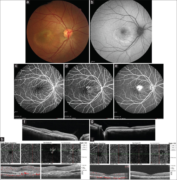
Figure 1.
(a) Fundus photograph of the right eye: Grey-white elevated retinal lesion with indistinct borders and retinal telangiectasia and scattered intraretinal hemorrhages in the temporal macula; (b) Fundus autofluorescence of the right eye shows a ring-shaped area of hyper autofluorescence with stippled central hypo autofluorescence compatible with location of choroidal neovascularization; (c-e) Fluorescein angiography of the right eye revealed a superior foci of early leakage in the macula of the right eye that through the late phases of angiography increased in size and borders faded gradually compatible with classic choroidal neovascularization and other inferior foci of stippled hyperfluorescence that was compatible with occult choroidal neovascularization; (f) Enhanced depth imaging optical coherence tomography of the right eye shows subretinal fluid and hyperreflective materials in the location of macular choroidal neovascularization with increased choroidal thickness especially in the nasal retina; (g) Enhanced depth imaging optical coherence tomography of the left eye shows relatively increased choroidal thickness with normal retinal structure; (h) Optical coherence tomography angiography confirmed the presence of choroidal neovascularization in the right eye; (i) Optical coherence tomography angiography of the right eye 1 month after 2 monthly intravitreal bevacizumab injections

