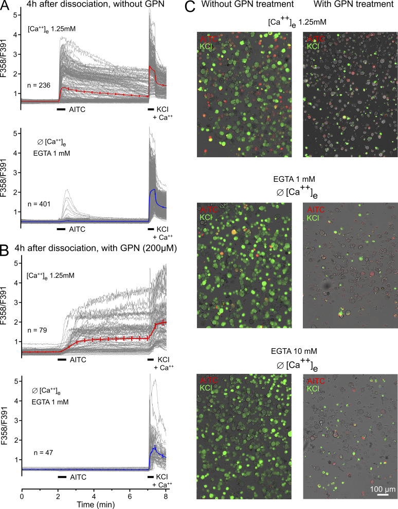
Figure S2.
Wide-field Fura-2 microfluorimetry data from freshly dissociated mouse DRG neurons. (A and B) Each panel represents pooled results from three to four measurements on two to three different days; n represents the mean number of viable (i.e., KCl-responsive) neurons per field of view. Captions indicate the extracellular calcium concentrations used and presence or absence of 1 mM EGTA; bars indicate the applications of 100 µM AITC and 60 mM KCl to activate TRPA1 and, by unspecific depolarization, voltage-gated calcium channels in the plasma membrane, respectively. (A) Naive DRG neurons without pretreatment, 35% responding with calcium transients to AITC in presence of extracellular calcium (upper panel), 14% in calcium-free condition (+ 1 mM EGTA); the bold and red- or blue-colored traces represent the mean (±SEM). (B) DRG neurons were pretreated with lysosome-disrupting GPN for 30 min during the Fura-2 exposure period in the presence of extracellular calcium, followed by 10-min GPN superfusion with calcium (upper panel) or calcium-free solution (+ 1 mM EGTA; lower panel). The yield of KCl-responsive neurons was strongly reduced compared with naive DRGs; the kinetics of AITC and KCl responses were reduced as well as their amplitudes; no neuron was activated by AITC in calcium-free solution; and the KCl responses were slowed and reduced. (C) Photomicrographs of false-color-stained AITC-responsive (red) and KCl-responsive (green) neurons; double-stained cells appear in orange/yellow, gray cells are unresponsive, which is the dominant color following GPN pretreatment (right column) irrespective of presence (upper row) or absence (lower rows) of extracellular calcium or of 1 or 10 mM EGTA, compared with naive DRG neurons (left column). Data from A and photomicrographs from C are also presented in Fig. 2, A and C.

