Figure 2.
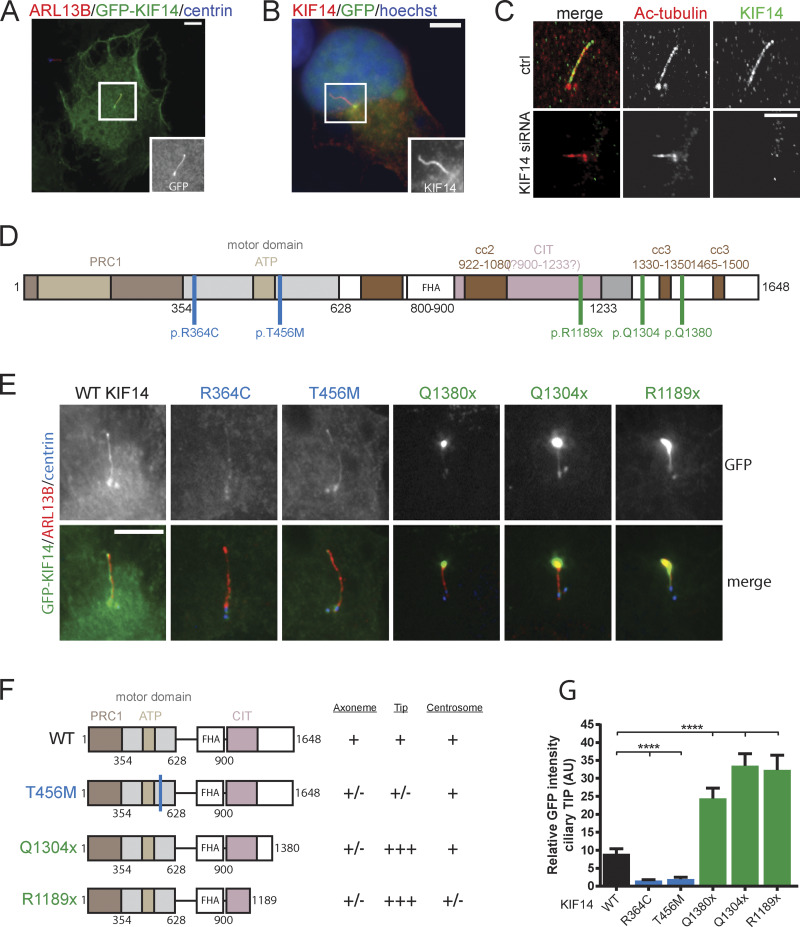
KIF14 localizes to primary cilia in interphase cells. (A) Representative image of IF staining detecting GFP-KIF14 (green) localization to the ciliary axoneme after overexpression in hTERT RPE-1, ARL13B (red), or centrin (blue); scale bar, 10 µm. (B) Representative image of IF staining detecting untagged KIF14 (red) localization to the ciliary axoneme after overexpression in hTERT RPE-1, GFP (green), or DNA (blue); scale bar, 10 µm. (C) Representative images of high-resolution microscopy analyses of endogenous KIF14 (green) localization to the ciliary axoneme and Ac-tub (red) in control vs. KIF14-depleted hTERT RPE-1 cells; scale bar, 5 µm. (D) Scheme of KIF14 mutants (Reilly et al., 2018) transfected into hTERT RPE-1 cells. PRC1, Protein Regulator of Cytokinesis 1 - binding domain; CIT, Citron Kinase - binding domain; FHA, forkhead-associated domain. (E) Representative images of IF microscopy analysis of KIF14 truncated mutant localization, ARL13B (red), GFP-KIF14 (green), and centrin (blue); scale bar, 20 µm. Note the accumulation of GFP-KIF14 in the ciliary tip upon transfection of C-terminally truncated mutants. (F) Graphical overview of GFP-KIF14 mutant localization. (G) Quantification of the GFP-KIF14 ciliary tip accumulation for different KIF14 mutants (relative fluorescent signal, arbitrary units). Asterisks indicate statistical significance determined using an unpaired t test, n = 2, N ≥ 55. AU, arbitrary units.

