Figure 3.
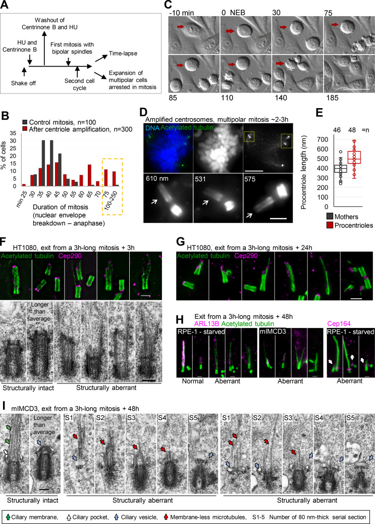
Prolonged mitosis leads to the formation of over-elongated and structurally and functionally aberrant centrioles. (A) A scheme delineating experimental design used to generate cells with amplified centrosomes leading to prolonged mitosis. (B) Quantification of duration of mitosis in control HeLa cells and HeLa cells containing amplified centrosomes. Cells were recorded by time-lapse in 5-min time intervals. (C) Example of a cell arrested in prometaphase for 140 min (red arrow). (D and E) Cells arrested in mitosis for ∼2–3 h were collected and analyzed by expansion. (D) Cell arrested in mitosis with multipolar spindle and over-elongated procentrioles (arrows). Numbers indicate procentriole length. (E) Quantification of centriole length in mitotically arrested cells. (F and G) Examples of aberrant centrioles analyzed by expansion and EM 3 h and 24 h after release from prolonged mitosis. (H) Representative examples of expanded structurally intact and aberrant centrioles/cilia 48 h after the release form a 3-h-long mitosis. ARL13B staining was used to distinguish normal looking from aberrant cilia. Arrows point to aberrantly localized Cep164. (I) Electron micrographs of structurally intact and aberrant centrioles in ciliation, 48 h after the release from prolonged mitosis. Scale bars, 20 µm in C; 20 µm for mitotic cell; 2 µm for enlarged centrioles in D, and for F, G, and H; 0.2 µm for EM in F and I.

