Figure 4.
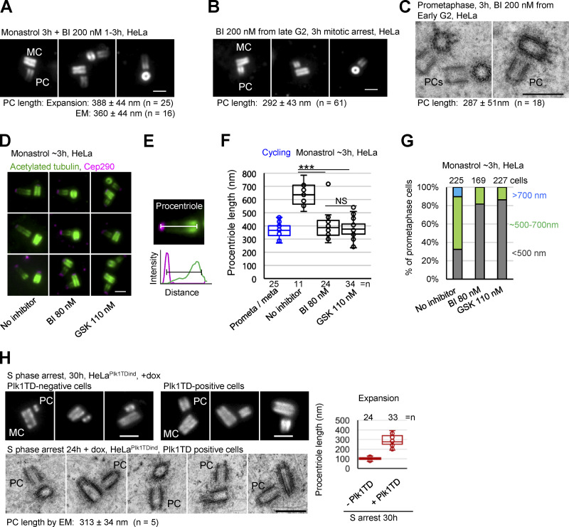
Plk1 is required for procentriole over-elongation in mitosis. (A) Expanded centrioles from cells arrested in mitosis for 3 h and treated with Plk1 inhibitor BI. (B) Expanded centrioles from cells arrested in mitosis by BI. (C) Electron micrographs of centrioles from cells arrested in mitosis by BI. (D) Expanded centrioles from cells arrested in mitosis and treated with Plk1 inhibitors BI and GSK. (E) Approach used to quantify procentriole length using acetylate tubulin and Cep290 signals. (F) Quantification of procentriole length using method in E. ***, P ≤ 0.001. (G) Quantification of procentriole phenotypes in expanded mitotically arrested cells. Procentrioles were scored for the length of their acetylated tubulin signals. Cells were placed in the green or blue category by the presence of at least one procentriole of indicated length. Cells with both procentrioles shorter than ∼500 nm were placed in the gray category. (H) Examples of centrioles from S phase arrested cells with/without active Plk1 (Plk1TD) analyzed by correlative conventional/expansion microscopy and EM. Plot: quantification of the length of procentriole acetylated tubulin signal from expanded samples. MC, mother centriole; PC, procentriole; average procentriole (PC) length, SD, and centriole number are indicated below images; scale bars, 0.5 µm for EM; 2 µm for expansion.

