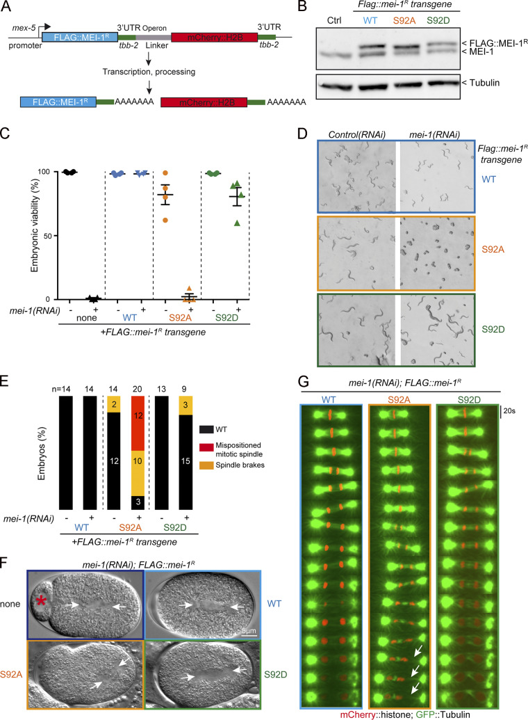
Figure 5.
The nonphosphorylatable MEI-1 S92A version is toxic in vivo in the absence of endogenous MEI-1. (A) Schematic showing the strategy used for expressing RNAi-resistant mei-1R S92A and mei-1RS92D transgenes (mei-1R) in the C. elegans germline. The operon linker is the intercistronic region from the gpd-2/gpd-3 operon. (B) Western blot analysis of embryonic extracts of the indicated genotype using MEI-1 (upper panel) and tubulin (lower panel) antibodies. (C) Viability of embryos expressing FLAG::mei-1R transgenes WT and mutants upon RNAi-mediated inactivation of endogenous mei-1. Error bars represent SEM; n = 4 independent experiments performed in triplicate with >100 embryos each. (D) DIC images of animal progeny with the indicated genotype exposed to control or mei-1(RNAi) for 36 h at 23°C. (E) Percentage of mitotic embryos similar to WT (black bars), presenting a misaligned mitotic spindle (red bars) or a mitotic spindle that breaks during mitosis (orange bars). The number of embryos analyzed is indicated and was generated by aggregation of more than three independent experiments. (F) DIC images of one-cell-stage embryos of the indicated genotype upon inactivation of endogenous mei-1. Arrowheads indicate the mitotic spindle. (G) Live images of mitotic spindles of the indicated genotype carrying GFP::α-Tubulin (green) and Histone2B::mCherry (red) transgenes. Arrows indicate locations where the mitotic spindle breaks.

