Figure 6.
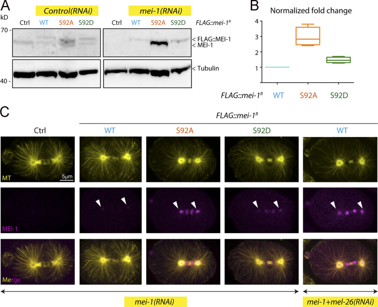
Site-specific MEI-1 phosphorylation (S92) is necessary and sufficient to target MEI-1 for degradation. (A) Western blot analysis of embryonic extracts from N2 (WT control, ctrl), FLAG::MEI-1 WT (light blue), FLAG::MEI-1 S92A (orange), and FLAG::MEI-1 S92D (green), animals exposed to mock (ctrl) or mei-1(RNAi) using MEI-1 (upper panel) and tubulin (lower panel, loading control) antibodies. (B) Quantification of the ratio of MEI-1 versus tubulin signal intensity over three independent experiments revealed that MEI-1 levels accumulate by threefold in FLAG::MEI-1 S92A embryos compared with the FLAG::MEI-1 WT control or FLAG::MEI-1 S92D upon endogenous inactivation of mei-1 by RNAi. Box-and-whiskers plot presenting MEI-1 levels quantified over three experiments. The whiskers represent the min and max values. (C) Representative spinning disk confocal micrographs of fixed early embryos of the indicated genotype immunostained with MEI-1 (magenta) and MT (yellow) antibodies. White arrowheads indicate the persistence of MEI-1 in mitosis at the centrosomes and the chromosomes. Additional representative images of embryos in metaphase are presented in Fig. S4.

