Figure S3.
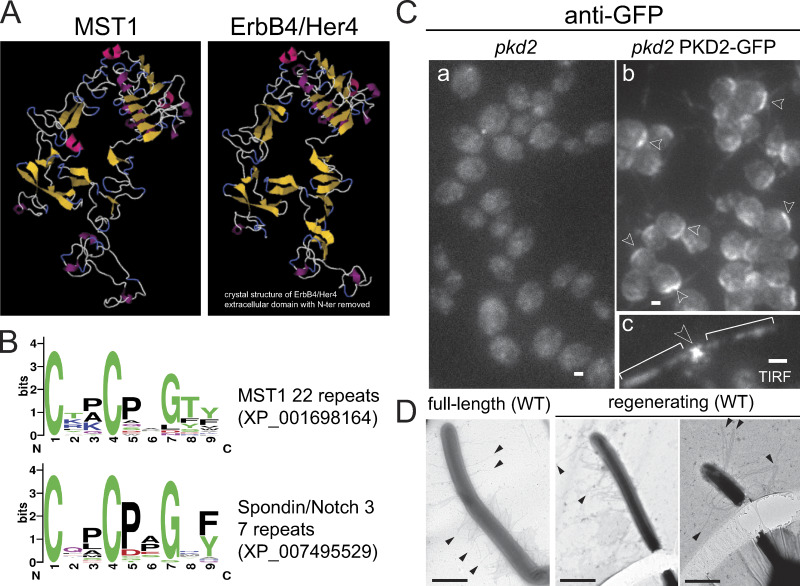
MST1 has an EGF-like extracellular fold. (A) Phyre2 homology-based structures of the C-terminal domain of MST1 and the extracellular domain of the receptor tyrosine-protein kinase ErbB4/Her4 showing the conservation of the EGFR extracellular domain fold (Kelley et al., 2015). (B) Weblogo of the 22 cysteine-rich repeats from MST1 and 7 similar motifs of the extracellular domain of Spondin/Notch3 (Crooks et al., 2004). (C) Staining of methanol-fixed pkd2 (a) and pkd2 PKD2-GFP (b) cells with anti-GFP. Arrowheads in b indicate PKD2-GFP signals near the basal bodies. TIRF live image of a pkd2 PKD2-GFP cell (c). Arrowhead in c indicates x-shaped localization of PKD2-GFP near the basal bodies. Brackets, cilia. Bar = 2 µm. (D) Whole-mount negative staining of wild-type cells showing mastigonemes of similar length on full-length and regenerating cilia. Arrowheads, mastigonemes. Bars = 1 µm.

