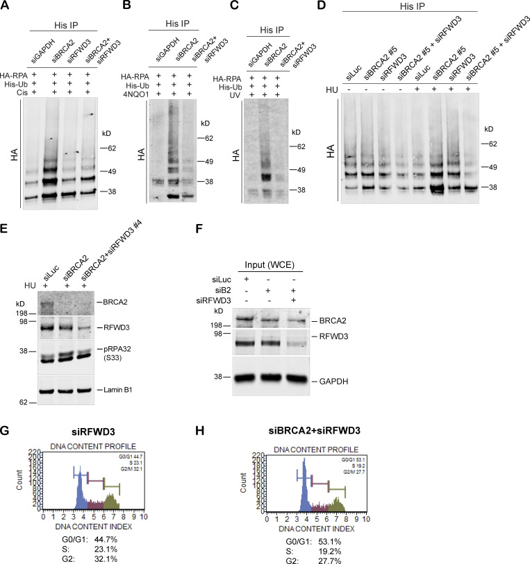
Figure S3.
Hyperubiquitination of RPA after BRCA2 depletion is performed by the E3 ligase RFWD3. (A–C) Immunoprecipitation analysis of RPA ubiquitination in HEK293T cells transfected with indicated siRNAs. Cells were treated with the following drugs: 2 µM cisplatin for 5 h (A), 1 mg/ml 4NQO1 for 3 h (B), or 30 J/m2 of UV for 3 h (C). (D) Immunoprecipitation analysis of RPA ubiquitination in response to HU in HEK293T cells treated with indicated siRNAs. A different BRCA2-specific siRNA (siBRCA2#5) was used for this experiment. (E) Western blot analysis of RPA32 accumulation after disrupting RPA ubiquitination by codepletion of RFWD3 in BRCA2-depleted cells. A different RFWD3-specific siRNA (siRFWD3#4) was used for this experiment. (F) Western blot analysis of whole-cell lysate served as input for iPOND. (G and H) Cell cycle analysis of RFWD3-depleted and BRCA2/RFWD3–codepleted cells by MUSE-based cell cycle assay. Related to Fig. 5.

