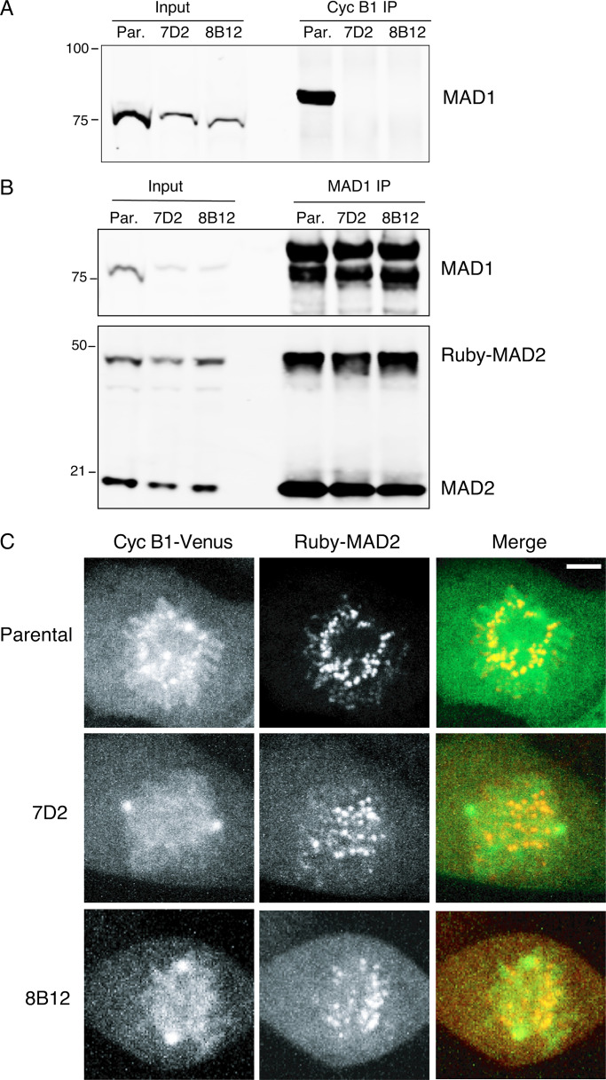
Figure 2.
The E53K/E56K mutation prevents MAD1 binding to cyclin B1 but not MAD2. (A) Parental RPE cyclin B1-Venus+/−:Ruby-MAD2+/− cells (Par.) or clones 7D2 and 8B12 carrying a homozygous mutation of E53/E56K in MAD1 were synchronized to enrich for G2 phase and mitosis; cyclin B1 (Cyc B1) was immunoprecipitated, subjected to SDS-PAGE, and immunoblotted with anti-MAD1 antibodies. (B) Parental RPE cyclin B1-Venus+/−:Ruby-MAD2+/− cells (Par.) or clones 7D2 and 8B12 were synchronized in G2 and M phase, and MAD1 was immunoprecipitated, and immunoblotted with anti-MAD1 (upper panel) and anti-MAD2 (lower panel) antibodies. (C) Parental RPE cyclin B1-Venus+/−:Ruby-MAD2+/− cells or MAD1 E53/E56K clones 7D2 and 8B12 were assayed by spinning disk confocal time-lapse microscopy. Images of maximum intensity projections of cyclin B1-Venus (left, green), Ruby-MAD2 (middle, red), and the merged image (right) for a representative prometaphase cell are shown. See Videos 1, 2, and 3. Data shown for all panels are representative of three independent experiments. Total number of cells: parental n = 21 cells, clone 7D2 n = 16 cells, clone 8B12 n = 10 cells. Scale bar, 3 µm.

