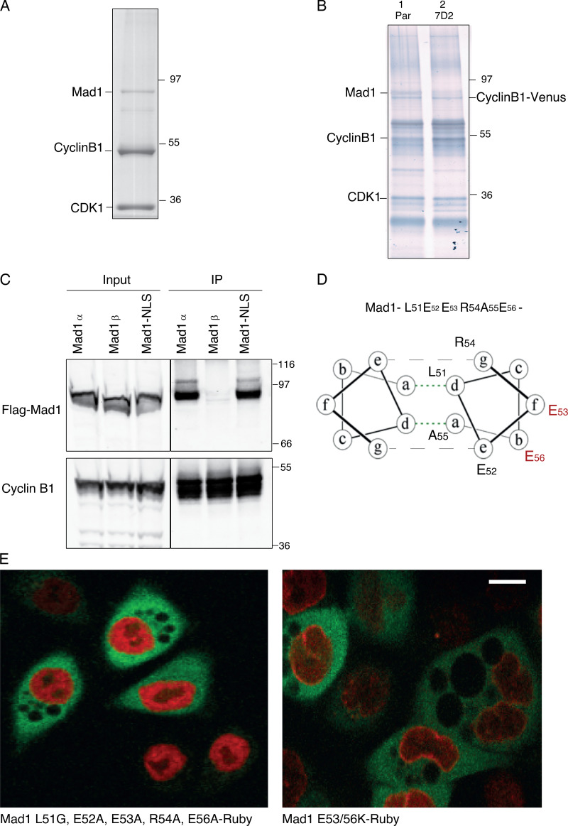
Figure S1.
Cyclin B1 binds to MAD1 through the acidic face of a helix encoded by exon 4. Related to Fig. 1. (A) Colloidal blue stained SDS-PAGE gel of cyclin B1 immunoprecipitated from HeLa cells. Marked bands were excised and identified by mass spectrometry. (B) Silver-stained SDS-PAGE gel of cyclin B1 immunoprecipitates from RPE cyclin B1-Venus+/−: Ruby-MAD2+/− cells (lane 1) and MAD1 E53K/E56K clone 7D2 (lane 2). (C) Cyclin B1 immunoprecipitates from HeLa cells expressing Flag-epitope tagged MAD1α or MAD1β or MAD1 with a mutated nuclear localisation sequence KKR79-82AAA (Mad1-NLS), probed with anti-FLAG (upper panel) or anti-cyclin B1 (lower panel) antibodies. (D) Heptad registration of acidic residues of MAD1 within coiled-coil configuration, predicted using PairCoil2. (E) Confocal image of HeLa cyclin B1-Venus+/− (green) cells transfected with either MAD1 L51G/E52A/E53A/R54A/E56A-Ruby (left panel, red) or MAD1 E53/56K-Ruby (right panel, red). Scale bar, 10 µm.

