Figure 1.
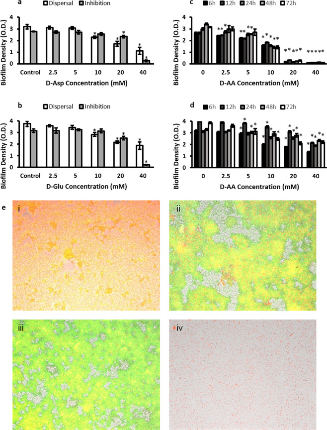
(a) Inhibition and dispersal of S. aureus biofilm using 2.5, 5, 10, 20 and 40 mM D-Asp. (b) Inhibition and dispersal of S. aureus biofilm using 2.5, 5, 10, 20 and 40 mM D-Glu. (c) Inhibition of S. aureus biofilm using equimolar concentrations of D-AA. (d) Dispersal of S. aureus biofilm using equimolar concentrations of D-AA. (a-d) One-way ANOVA showed an overall significant difference within the data and was followed by a post-hoc t-test with Bonferroni correction to see which concentration significantly (p < 0.0083 was taken as significant; indicated by *) significantly inhibited or dispersed biofilms; n = 3, ±S.D. (e) Fluoresence imaging showing 72 h biofilm formation. (i) Control (ii) inhibited with 40 mM D-Asp (iii) inhibited with 40 mM D-Glu (iv) inhibited with equimolar concentrations of D-Asp and D-Glu (40 mM of each); D-Asp = D-Aspartic acid; D-Glu = D-Glutamic acid; D-AA = D-Asp and D-Glu.

