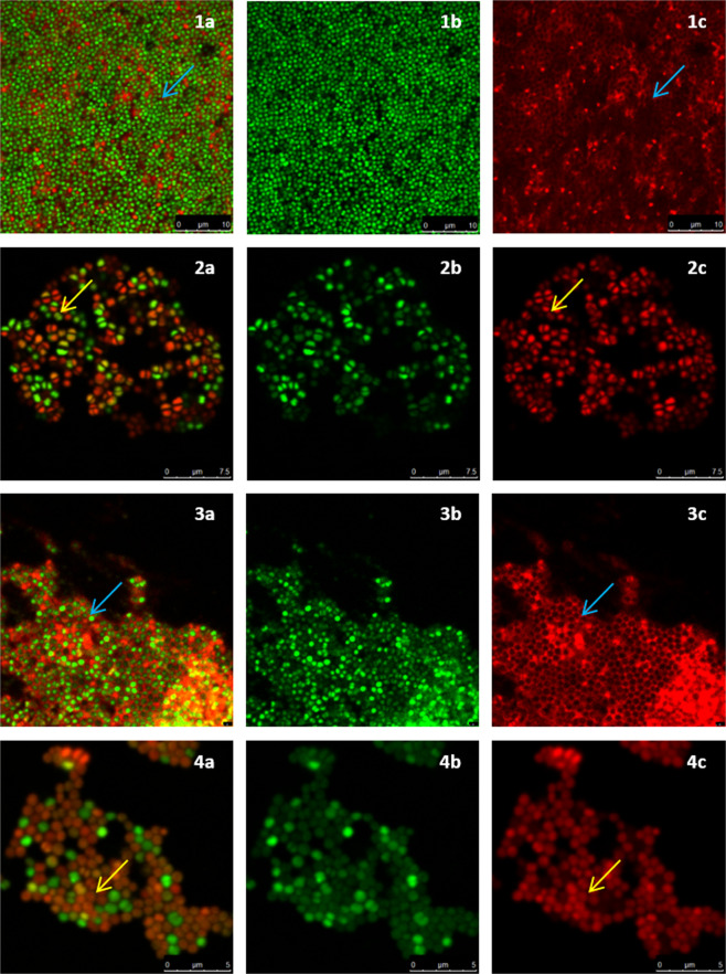
Figure 5.
Confocal images of inhibited S. aureus biofilms. (a) combined channel; (b) SYTO 9 channel; (c) Promidium Iodide channel; (1) control biofilm; (2) inhibited with 40 mM D-AA (40 mM D-Asp and 40 mM D-Glu); (3) treated with Cip; (4) treated with 40 mM D-AA and 8 MLC Cip; blue arrow) presence of intercellular eDNA; yellow arrow) lack of intercellular eDNA. (1) Untreated S. aureus biofilms are reinforced by an organised honeycomb like meshwork made of interconnected intercellular eDNA. eDNA forms a filamentous mesh like structure within the biofilm and surrounds all cells. (2) Biofilms inhibited with D-AA show a complete lack of eDNA. (3) Biofilms inhibited by Cip express a lower population of S. aureus cells. A dense eDNA meshwork provides structure to these persisting cells. (4) Biofilm treated with a combination of D-AA and 8 MLC Cip lack in eDNA meshwork structure; D-AA = 40 mM D-Aspartic acid and 40 mM D-Glutamic acid; x MLC = x times minimum lethal concentration; Cip = ciprofloxacin.

