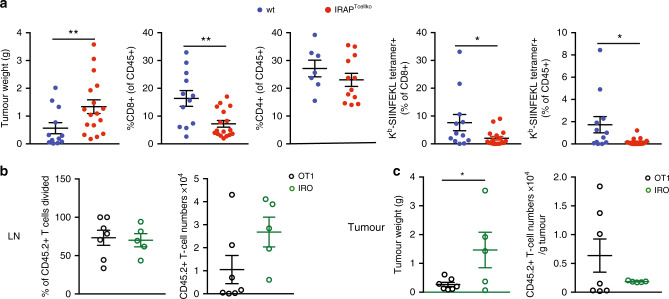
Fig. 7. IRAP is required for anti-tumour T-cell response.
a Wt or IRAPTcellko mice were injected s.c. with EG7-ova cells, and 12–14 days later, tumour weight and CD8+ T-cell response in the tumour were measured. Each dot represents a mouse. Values represent mean ± SEM of pooled results from three independent experiments. (Wt: n = 13 (except for graph, 3 where n = 7, IRAPTcellko: n = 16 (except for graph 3, where n = 12). Graphs 1: **p = 0.0041, 2: **p = 0.0018, 4: *p = 0.0466, 5: *p = 0.0247). b, c CD45.1 C57BL6 mice were injected s.c. with EG7-ova cells, and 10–11 days later, 3 × 10⁶ CTV-labelled OT1 or IRO T were injected i.p. The divisions of transferred T cells and their absolute numbers were evaluated after 6 days in the lymph nodes (LN) (b). The tumour mass and the numbers of T cells infiltrating the tumour were analysed 6 days after T-cell transfer (c). Each dot represents a mouse. Values represent mean ± SEM of pooled results from two independent experiments (OT1: n = 7, IRO: n = 5, *p = 0.0440). All p-values were calculated with two-tailed unpaired t tests. For additional information, see Supplementary Fig. 8.

