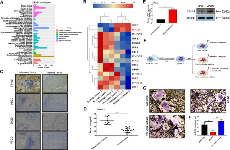
Fig. 1. IFN-λ1 was higher expressed from dendritic cells during inflammatory statement.
a The genes expression in immune system were analyzed by KEGG pathway enrichment analysis between chronic osteomyelitis-infected samples and non-infectious fractures samples. b Profiling of differentially expressed interferon-related genes between chronic osteomyelitis-infected samples and non-infectious fractures samples. c Representative images of immunohistochemical staining with monoclonal antibodies against IFN-λ1, CD80, CD86, and CD1a. d ELISA for IFN-λ1 concentrations from the serum of chronic osteomyelitis patients and non-infectious fracture patients. e Relative expression of IFN-λ1 on mRNA and protein level between LPS (100 ng/ml) pretreatment with dendritic cells and vehicle for 72 h. β-actin and GAPDH were used as an internal control. f Schematic representation of the experimental design of the establishment the co-culture between osteoclast and LPS-induced dendritic cells. g Representative images of TRAP staining of RAW264.7 cells treated with different groups. h Quantification of multinucleated TRAP-positive osteoclast number per well. The data in the figures represent the averages ± SD. Scale bars = 200 μm. Significant differences are indicated as *p < 0.05 or **p < 0.01 paired using Student’s t-test unless otherwise specified.

