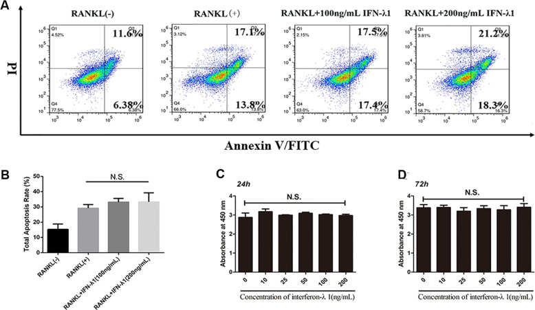
Fig. 3. IFN-λ1 could not make cytotoxicity during osteoclastogenesis.
a Flow cytometry analysis of the apoptosis rate of RAW264.7 cells treated with RANKL (100 ng/ml) and M-CSF (50 ng/ml) for 72 h with various doses of IFN-λ1. b Quantitative analysis of total apoptosis rate during osteoclastogenesis. c, d CCK-8 was performed in triplicate to analyze the cell viability of BMMs treated with varying doses of IFN-λ1 for 24 and 72 h with or without RANKL (100 ng/ ml) and M-CSF (50 ng/ml). The data in the figures represent the averages ± SD. N.S. represented as no significant difference. Significant differences are indicated as *p < 0.05 or **p < 0.01 paired using Student’s t-test unless otherwise specified.

