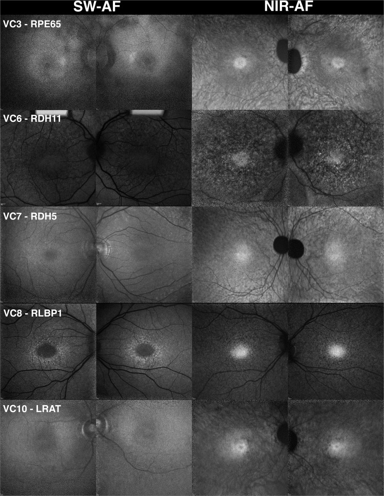
Figure 1.
Fundus autofluorescence presenting in patients exhibiting mutations in visual cycle genes (VC). The short-wavelength autofluorescence (SW-AF) images of five patients carrying mutations in visual cycle genes, including RPE65, LRAT, RLBP1, RDH5, and RDH11, indicate that the feature common to this cohort is dark or absent autofluorescence illustrated by fluorescence intensity in the vasculature and optic nerve that is similar to the rest of the macula. Choroidal vessels are visible in the near-infrared autofluorescence (NIR-AF) images in the patients with mutations in RPE65, LRAT, and RDH5. This suggests a reduction in NIR-AF from RPE melanin. A hyperautofluorescent ring can be seen in the patient with mutations in RLBP1 by short-wavelength autofluorescence. All other patients do not demonstrate hyperautofluorescent rings in either short-wavelength or near-infrared autofluorescence images.

