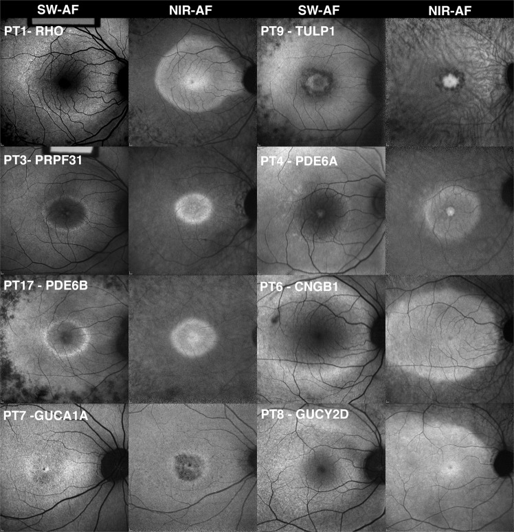
Figure 2.
Fundus autofluorescence images obtained from patients with mutations in phototransduction genes (PT). The short-wavelength autofluorescence (SW-AF) and near-infrared autofluorescence (NIR-AF) images obtained from patients with mutations in phototransduction genes (RHO, PRPF31, PDE6B, GUCA1A, TULP1, PDE6A, CNGB1, and GUCY2D), exhibit hyperautofluorescent rings in both SW-AF and NIR-AF images. The exception is the patient with a mutation in GUCY2D, who presents with a sectoral hyperautofluorescent arc in the superior retina. In all cases, autofluorescence was sufficient to enable imaging and the macula appeared brighter than the ophthalmic vessels and the optic nerve.

