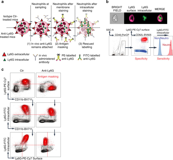
Fig. 1. Intracellular Ly6G staining overcomes surface Ly6G unavailability.
a Schematic representation of the antigen masking related issue. Upon anti-Ly6G treatment, the in vivo delivered antibody remains bound to the surface antigen even after sampling, impairing fluorochrome-labeled additional binding. In contrast, the intracellular antigen remains available. b (top) Representative image acquired with ImageStream of a neutrophil where the Ly6G antigen has been successively and specifically stained on the membrane then in the intracellular compartment. (down) Representative flow cytometry analysis emphasizing the specificity and sensitivity of the intracellular Ly6G staining. c Mice were treated for 24 h with an isotype control (left) or anti-Ly6G (right) antibody, after which the bone marrows were sampled. CD45+CD11b+Ly6Gintra+ neutrophils are shown in red. In the anti-Ly6G-treated group, cells were not detectable using the extracellular staining because of the antigen masking.

