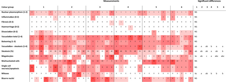
Table 1.
Histological observation of lumpfish livers of different colours. Individual fish score (nuclear pleomorphism, inflammation, fibrosis, haemorrhage, dissociation, total vacuolation, ballooning and steatosis), percentage (steatosis (%)) or number of cells at high-power field 10 (megalocyte, multinucleated, necrotic/apoptotic, mitoses and bizarre nuclei cells). Colour is proportional to values. Different letters indicate significant differences (ANOVA, post-hoc Tukey’s, P < 0.05, or χ2-test, P < 0.05) between liver colours. NS indicates no significant differences.

