
Figure 2.
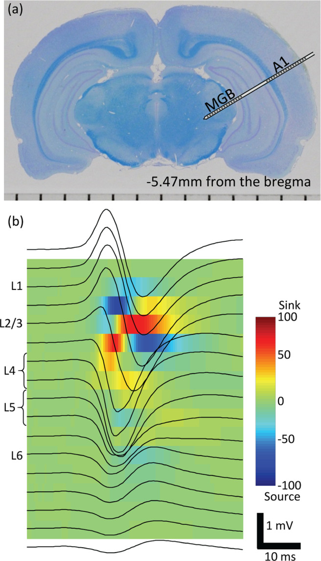
Electrophysiological experiments. (a) Recording with microelectrode array in the A1 and MGB. A coronal histological section of a representative test animal. (b) Laminar recording and current source density analysis in the A1. Upon recording the AEPs across A1 layers (black traces on the image), the CSD analysis was performed to locate the test sites in L1, L2/3, L4, L5 and L6. A1, primary auditory cortex; MGB, thalamus; AEP, auditory evoked potential; CSD, current source density; L, layer.