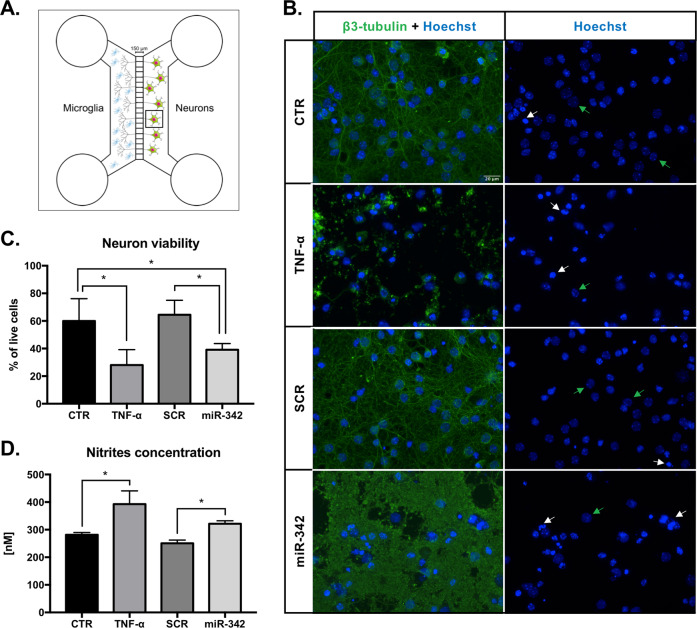
Fig. 7. miR-342 overexpression in microglia induces neurotoxicity.
a Neurons were cultured in PDL-coated coverslips previously attached to the Axon Investigation System. At day 13 of neuronal culture, transfected, TNF-α, or non-stimulated N9 microglia were added to the respective system, in direct contact with axons for 24 h. b Immufluorescence images of neurons after co-culture with N9 microglia. Left panel shows neurons stained with anti-β3-tubulin (anti-Alexa 488, green) and Hoechst (blue). Right panel shows only Hoechst nuclear staining used for neuron viability evaluation. White arrows highlight the nucleus of dead neurons, whereas green arrows highlight the nucleus of healthy neurons. Scale bar: 20 µm. c Neuron viability was addressed after counting the number of living and dead cells of 10 images per condition (mean ± SD, n = 4). d Co-cultures’ supernatants were collected for nitrite levels quantification using Griess reagent (mean ± SD, n = 4). Statistical significance: *p < 0.05 and **p < 0.01, Friedman test followed by Dunn’s multiple comparisons test.

