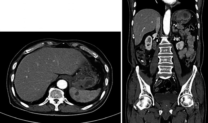
Fig. 2.
Stomach computed tomography shows an approximately 5.6 cm sized endoluminal protruding mass with delayed enhancement and area of necrosis in the posterior wall of gastric fundus, which is perigastric fat infiltration, abutting to the spleen and ill-defined wedge-shaped low attenuated lesion in the spleen.

