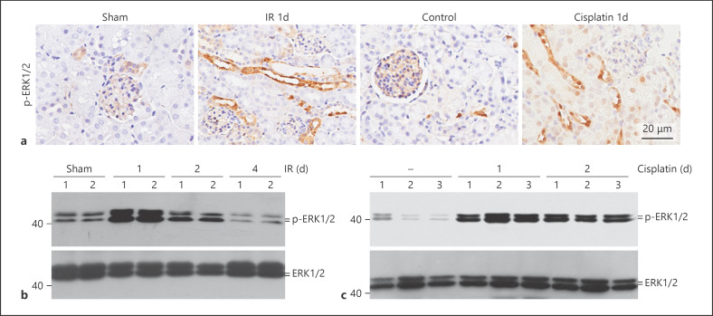
Fig. 2.
Erk1/2 signaling is activated in the kidneys with AKI. a Representative immunohistochemical staining images for p-Erk1/2 in kidneys at day 1 after IR or cisplatin injection, showing the activation of Erk1/2 signaling in tubular cells. b, c Western blot analysis showing the abundance of p-Erk1/2 in kidneys at different time points after IR (b) and cisplatin injection (c). The number indicates the individual animal.

