Fig. 5.
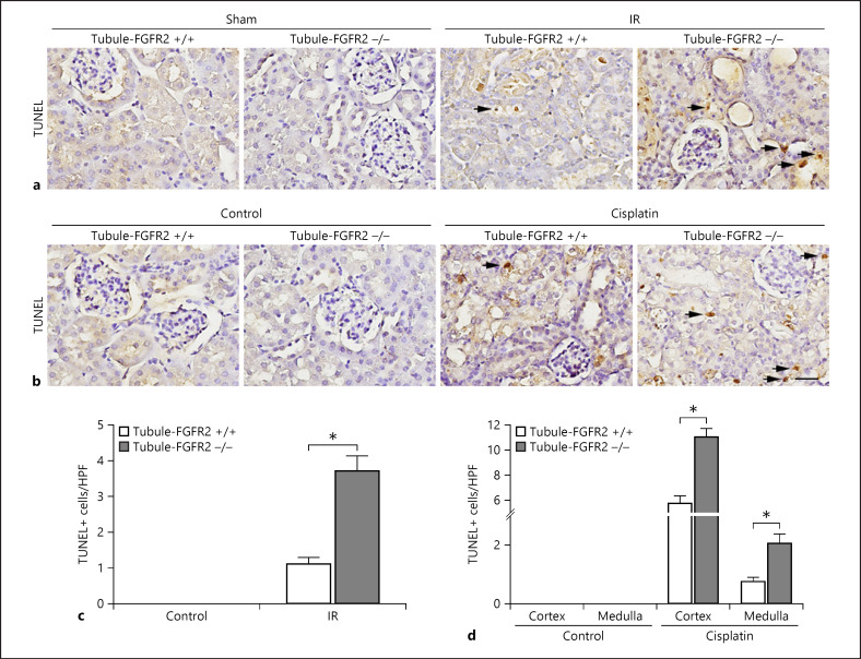
Tubular cells are more susceptible to apoptosis in mice with tubule-specific ablation of FGFR2. a, b Representative micrographs showing TUNEL staining for apoptotic cells in Tubule-FGFR2−/− kidneys and control littermates at day 2 after IR (a) and at day 3 after cisplatin injection (b). Black arrows indicate TUNEL staining-positive cells. Scale bar, 20 μm. c, d Quantitative data showing the TUNEL staining-positive cells in Tubule-FGFR2−/− mice and control littermates after IR (c) and cisplatin injection (d). Data are presented as TUNEL staining-positive cells per high-power field (HPF, 400×) in cortex and medulla. * p < 0.05 vs. control littermates after IR or cisplatin injection, n = 3.

