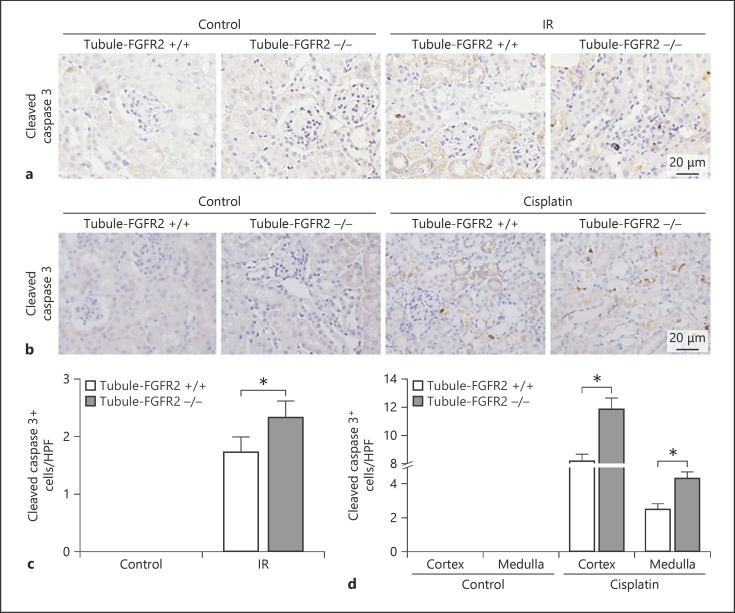
Fig. 6.
Ablation of FGFR2 aggravates caspase 3 cleaved in mice after cisplatin injection. a, b Representative micrographs showing the immunohistochemical staining of cleaved caspase 3 in Tubule-FGFR2−/− kidneys and control littermates at day 2 after IR (a) and at day 3 after cisplatin injection (b). c, d Quantitative data showing the cleaved caspase 3-positive cells in Tubule-FGFR2−/− mice and control littermates after IR (c) and cisplatin injection (d). Data are presented as TUNEL staining-positive cells per high-power field (HPF, 400×) in cortex and medulla. * p < 0.05 vs. control littermates after IR or cisplatin injection, n = 3.

