
Figure 1:
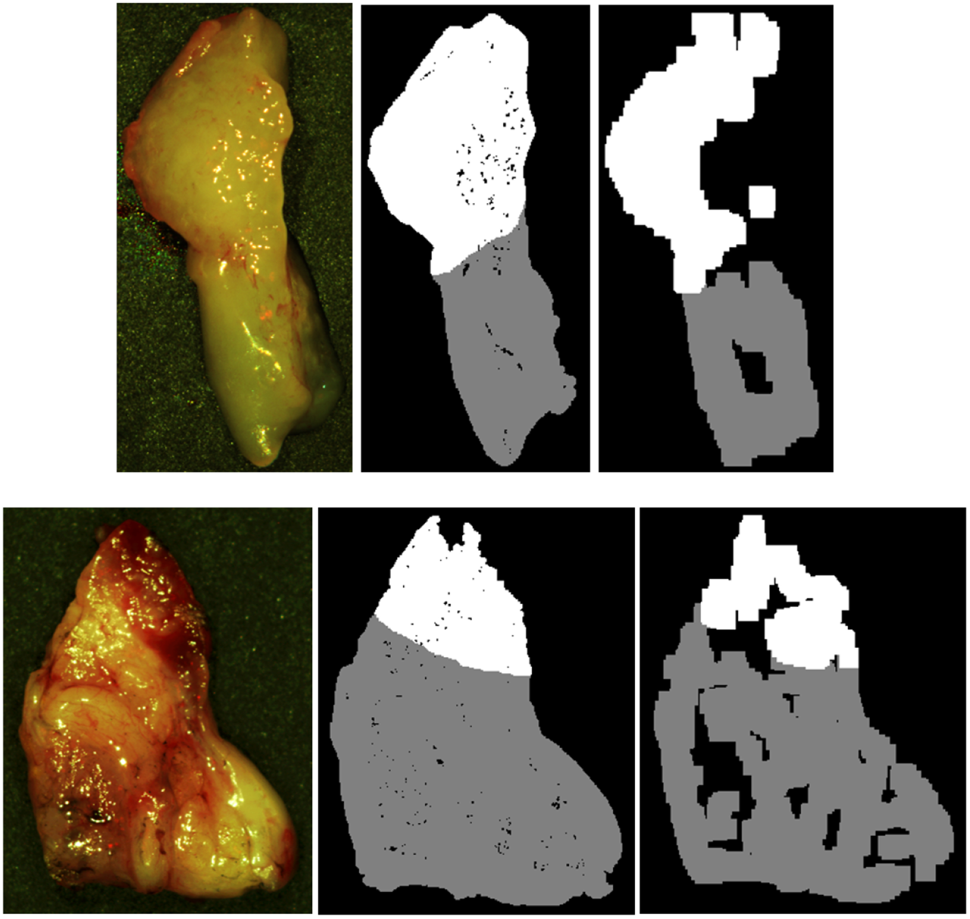
Two representative tissue specimens from different patients. Left to right: HSI-RGB composite image; binary ground-truth mask including glare regions, generated by only removing patches centered on specular glare; binary ground-truth mask avoiding glare regions, generated by sufficient area to extract 25×25 patches and avoiding specular glare. SCC in shown in white, and normal tissue shown in grey.