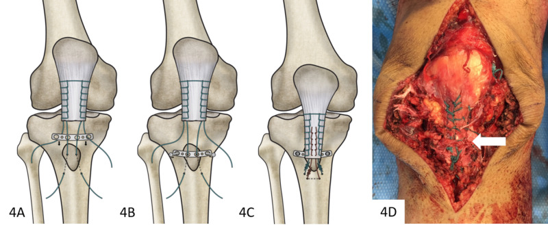
Figure 4. Sequence of plate fixation with final intraoperative appearance.
(A) A slotted plate was positioned after transosseous suture placement through tibia, tubercle fragment, and patellar tendon. (B) The plate was then secured atop the tubercle fragment. (C) Primary sutures were tensioned and a third, defunctioning suture (red) was placed through a distal pilot hole and secured to the patellar tendon with a running-locking technique. (D) Completed fixation with the tubercle fragment reduced, plate secured (white arrow, not visible), and the tendon reduced.

