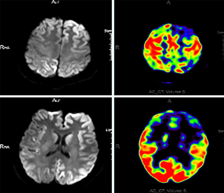
Fig. 4.
PET-MRI images of the same sCJD patient. Two images of PET-MRI scans in the same patient showing decreased FDG uptake in the cerebral cortex and basal ganglia. PET-MRI, positron emission tomography-magnetic resonance imaging; sCJD, sporadic Creutzfeldt-Jakob disease; FDG, fluorodeoxyglucose.

