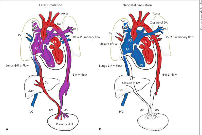
Fig. 3.
Schematic of normal fetal and neonatal cardiovascular circulation. a During fetal development, oxygenated and nutrient-rich fetal blood from the placenta passes to the fetus via the umbilical vein. Approximately half of this blood bypasses the liver via the DV and enters the IVC. The remainder enters the portal vein to supply the liver with nutrients and oxygen. Blood entering the RA from the IVC bypasses the RV as the lungs are not yet functioning, and then enters the LA via the FO. Blood from the SVC enters the RA, passes to the RV, and moves into the PA trunk. Most of this blood enters the aorta via the DA, a right-to-left shunt. The partially oxygenated blood in the aorta returns to the placenta via the paired UA that arise from the internal iliac arteries. b Under normal physiological conditions, when pulmonary respiration begins at birth, pulmonary blood pressure falls, causing blood from the main PA trunk to enter the left PA and right PA, become oxygenated at the lungs, and then return to the LA via the PV. The FO and DA close, eliminating the fetal right-to-left shunts. The pulmonary and systemic circulations in the heart are now separate. As the infant is separated from the placenta, the UAs occlude (except for the proximal portions), along with the umbilical vein and DV. Blood to be metabolized now passes through the liver. DV, ductus venosus; UV, umbilical vein; IVC, inferior vena cava; RA, right atrium; RV, right ventricle; LA, left atrium; FO, foramen ovale; SVC, superior vena cava; PA, pulmonary artery; DA, ductus arteriosus; UA, umbilical arteries; PV, pulmonary veins.

