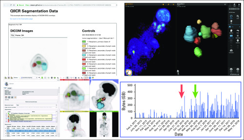
FIG 2.
Example demonstration of image analysis results interoperability enabled by DICOM. From bottom left corner clockwise, examples of platforms visualizing the same Digital Imaging and Communications in Medicine (DICOM) positron emission tomography (PET) segmentation dataset from the public QIN-HEADNECK collection20,61: 3D Slicer8 (free, open-source desktop application), OHIF Viewer38 (free, open-source Web viewer), Brainlab SmartBrush (commercial Food and Drug Administration–approved tumor outlining application). Results were collected as part of the DICOM4QI (DICOM for Quantitative Imaging) demonstration and connectathon organized by Quantitative Imaging Informatics for Cancer Research (QIICR) at the annual Radiologic Society of North America meeting since 2015.36,62 The histogram shows the significant increase in the The Cancer Imaging Archive–reported usage of the QIN-HEADNECK collection after the publication of the preprint in Nov 2015 (red arrow) and peer-reviewed paper in May 2016 (green arrow),20 which introduced imaging-related DICOM data (segmentations, measurements, clinical data) to accompany the imaging dataset.

