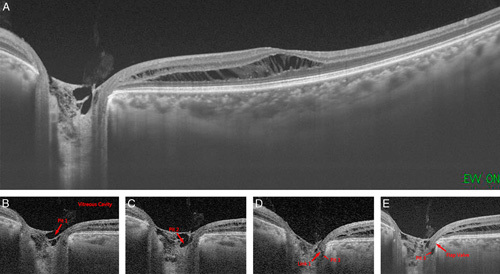
FIGURE 3.

A, Postoperatively optical coherence tomography images revealed posterior vitreous detachment, retinal edema, and retinoschisis ranging from the optic disc to the macula. B–E, Vitreous cavity was connected to the retinoschisis through pits 1 to 3 and a valve/flap-like structure. Arrow indicates the pits and the links between them.
