
FIG. 3.
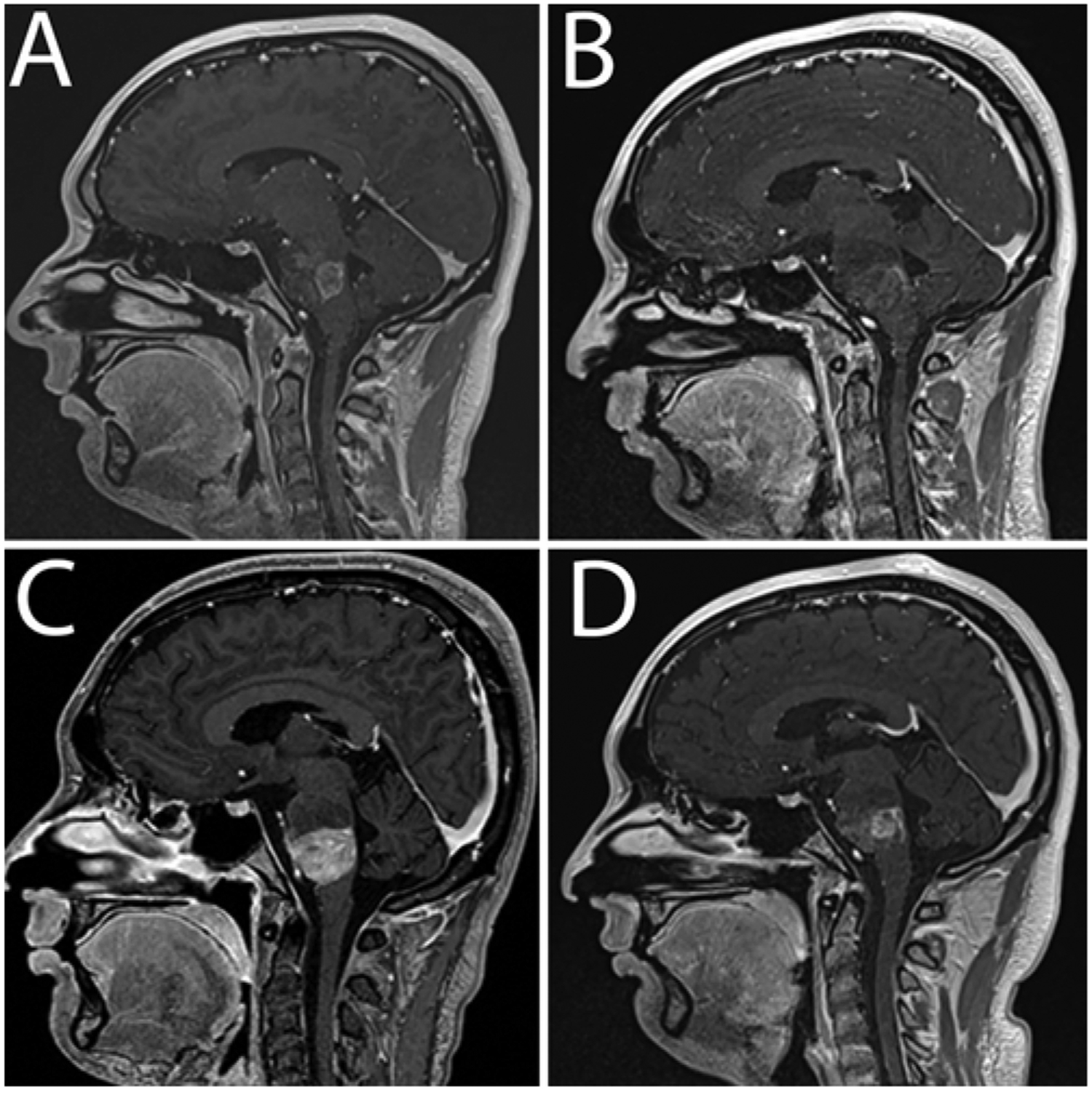
Patient 5. Sagittal contrast-enhanced T1-weighted MR images obtained preoperatively (A) and 1 month (B) and 2 (C) and 3 (D) months postoperatively. The 1-month follow-up image (B) shows minimal enhancement, low perfusion, and necrosis at the infusion site consistent with treatment response.