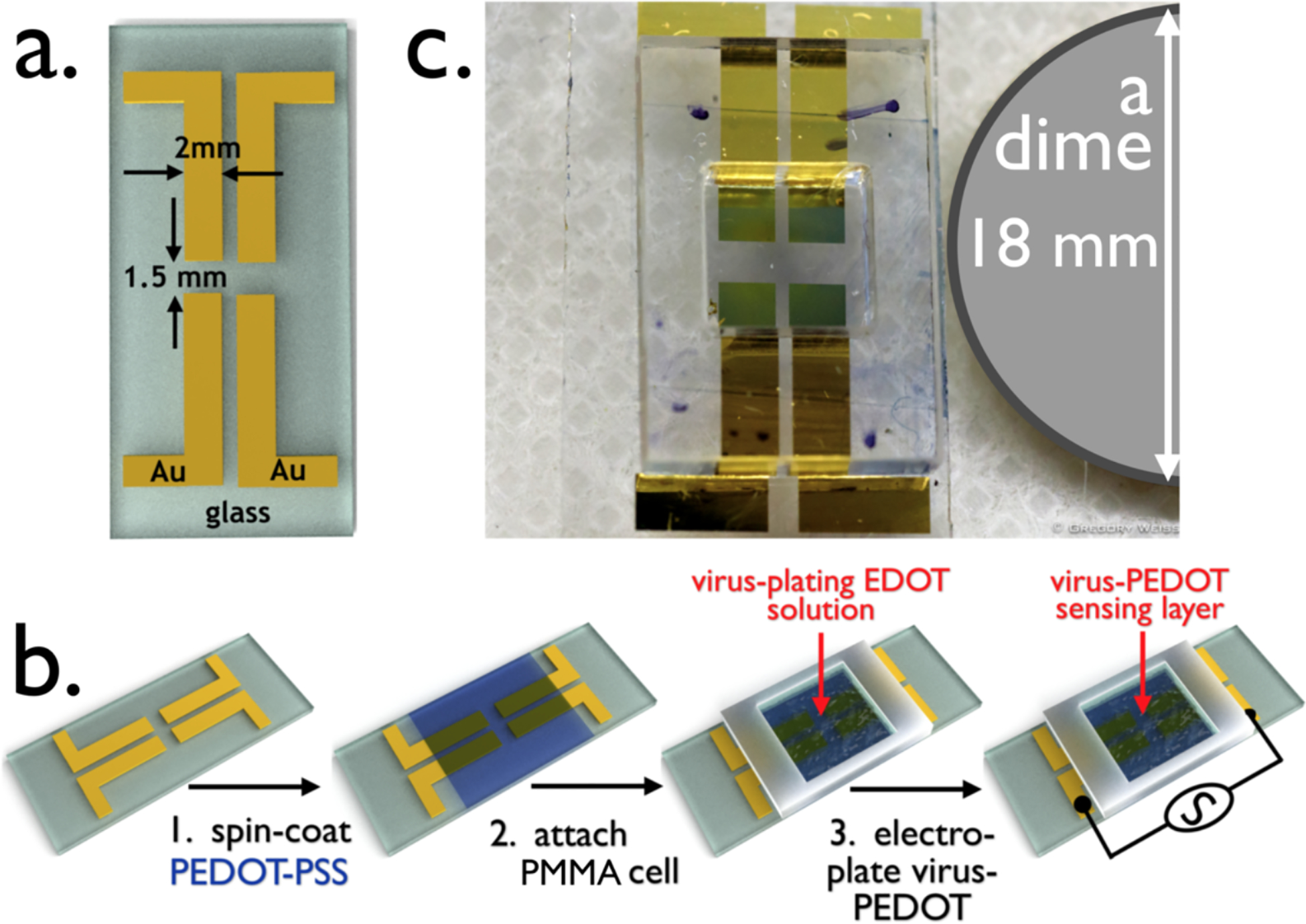
Figure 2.

The Virus BioResistor (VBR). a) Rendering of gold electrodes for a two-VBR chip showing its dimensions. The two electrodes at left comprise one VBR and the two on the right a second VBR. These two VBRs will share a single bioaffinity layer. b) The three-step process for fabricating a VBR: Step 1 – a conductive PEDOT-PSS base layer is spin-coated onto the gold-on-glass template shown in (a). This film is baked at 90 °C for 60 min; Step 2 – A poly(methylmethacrylate)(PMMA) cell is attached on top of the dried PEDOT-PSS film; Step 3 – the PMMA cell is filled with aqueous EDOT-virus plating solution, and a virus-PEDOT film is deposited by electrooxidation. This VBR biosensor is ready for use. c) Photograph of a two-VBR chip with PMMA solution cell.
