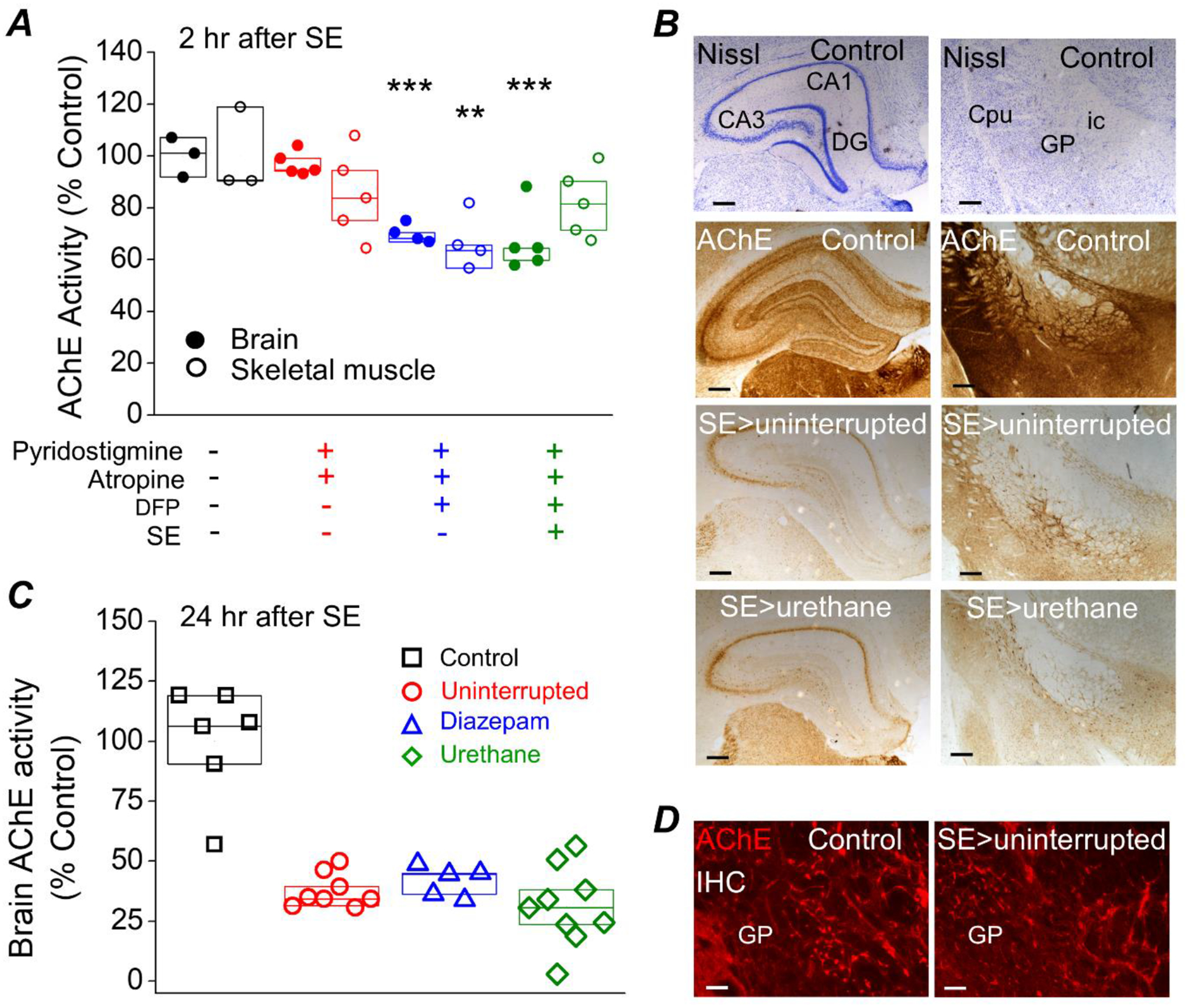
Figure 10. Acetylcholinesterase activity is unaltered by urethane or diazepam.

A, AChE inhibition in rat brain and skeletal muscle (from the hindleg, Vastus Lateralis) measured by an acetylcholinesterase assay is significantly reduced measured at 2 hours after SE in rats administered DFP. ** = p < .01, *** = p < .001, one-way ANOVA with posthoc Dunnett’s compared to the “no treatment” controls. Data are box plots with a 25 and 75 range. The symbols represent each individual rat within the group. B, the brain of a non-seizure control rat, a rat that experienced uninterrupted SE and a rat that was administered a single injection of urethane (0.8 mg/kg) 1 hour after SE was rapidly removed and bisected longitudinally at 24 h. One hemisphere was processed for immunohistochemistry. Coronal sections were stained for Nissl and acetylcholinesterase activity using the protocol described by Paxinos et al. (1980). The dark purple indicates the presence of the Nissl bodies and the brown precipitate is indicative of areas of high AChE activity. The top 2 images show the results of Nissl and the bottom 6 images reveal AChE activity in the hippocampus on the left and the globus palladus (GP), internal capsule (ic) and caudate putamen (Cpu) (25x total magnification) on the right. The Nissl and AChE activity stained sections were obtained from the same rats. Scale bar, 100 μm. C, inhibition of acetylcholinesterase in rat forebrain 24 h after DFP-induced SE was determined using an AChE activity assay and protein lysates obtained from half brains. AChE inhibition was similar regardless of whether the rats received diazepam or urethane. The controls were non-seizure rats that were administered pyridostigmine bromide, ethylatropine bromide and water instead of DFP. Data are box plots with a 25 and 75 range. The symbols represent each individual rat within the group. p > .05, one-way ANOVA with posthoc Bonferroni (diazepam vs. urethane). D, immunohistochemistry for AChE was performed on coronal sections revealing positive fluorescent staining of AChE in the globus pallidus (100x total magnification) indicating the presence of acetylcholinesterase (bright red staining). Scale bar, 30 μm. IHC= immunohistochemistry. CA1, cornu ammonis 1; CA3, cornu ammonis 3; DG, dentate gyrus.
