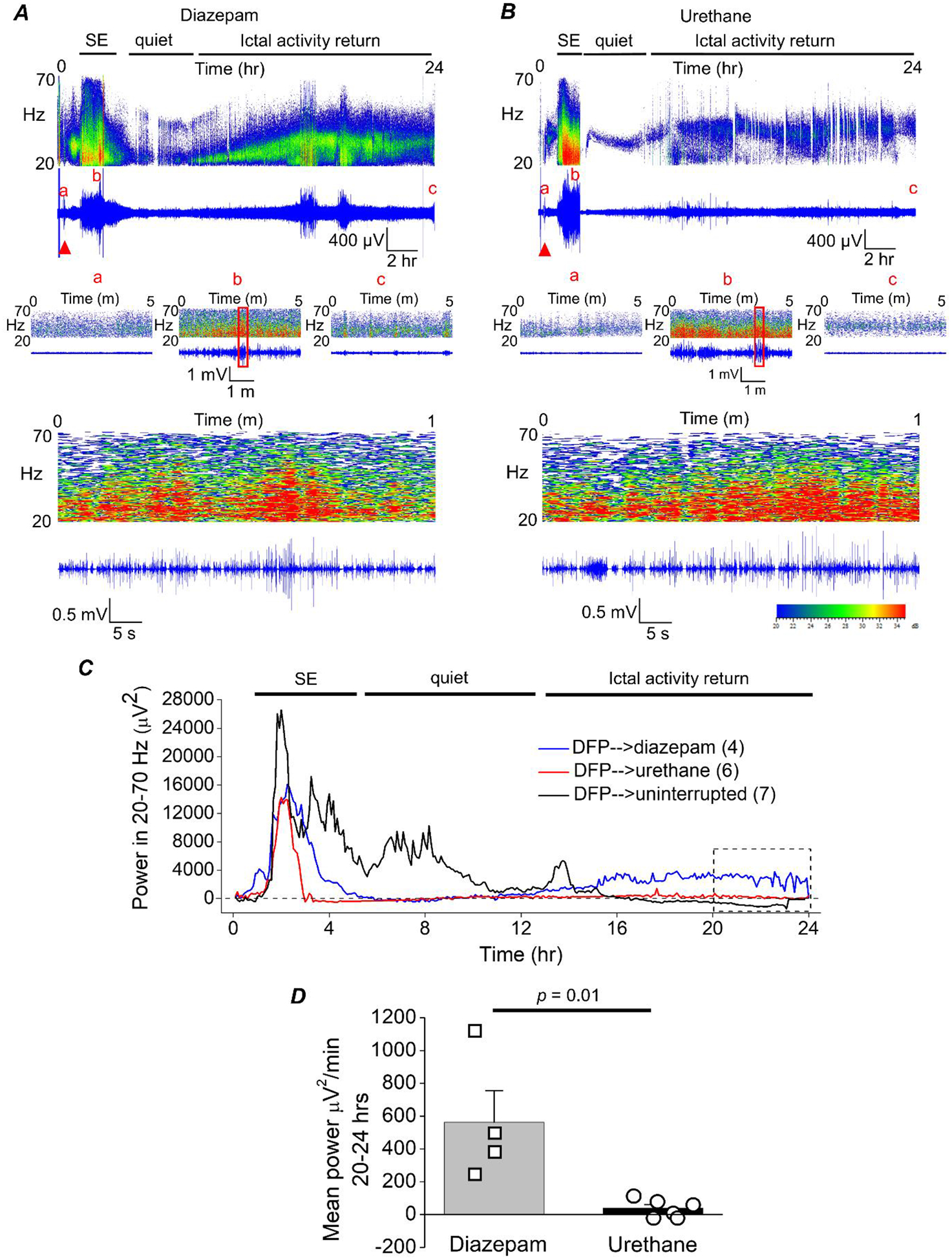
Figure 3. The return of seizure activity following 1 hour of DFP-induced SE is suppressed by urethane.

A, cortical electroencephalography (EEG) activity was recorded prior to and during SE induced by exposure to DFP for 24 h. A representative EEG trace and sonogram from the cortical recording of an adult male rat showing increased spike activity just after exposure to DFP (red triangle) that develops into SE (denoted by the bar at the top). Spike activity was quieted by administration of diazepam (10 mg/kg, ip) 1 h after SE onset, but returned within a few hours as denoted by the ictal activity return phase. Above each raw EEG trace is a sonogram of the spike activity obtained in Spike2. The colors of the sonogram indicate the spectral power density in decibels (dB) at the indicated frequency. The middle panels show magnified 5 min intervals of the recording taken from the baseline prior to DFP (a), at the peak intensity of SE (b) and at the end of the recording (c). The area within the red outlined box in panel b was magnified and shown in the bottom panel that represents a 1 min interval of the recording during SE. B, a representative EEG trace from the cortical recording of an adult male rat shows increased spike activity just after exposure to DFP (red triangle) that developed into SE, which was quieted following brief exposure to isoflurane (inhaled) and subsequent injection of urethane (0.8 g/kg, sc) 1 h after SE onset. C, the EEG power in the 20–70 Hz bandwidth averaged over 300 sec epochs during the 24 h period for 4 diazepam-treated, 6 urethane-treated rats and 7 rats that experienced uninterrupted SE. The dashed line indicates baseline power before DFP administration. D, a significant difference was detected between the two treatment groups [diazepam (n = 4) and urethane (n = 6)] in the EEG power in the 20–70 Hz bandwidth using power analysis during the final 4 h of the EEG recording (area within the dashed box in panel C). Data are the mean ± standard error of the mean. p = .01, student’s t-test. The symbols represent each individual rat within the group.
