
Figure 5. Neuroprotection by urethane in nuclei of the ventral temporal lobe following DFP-induced status epilepticus.
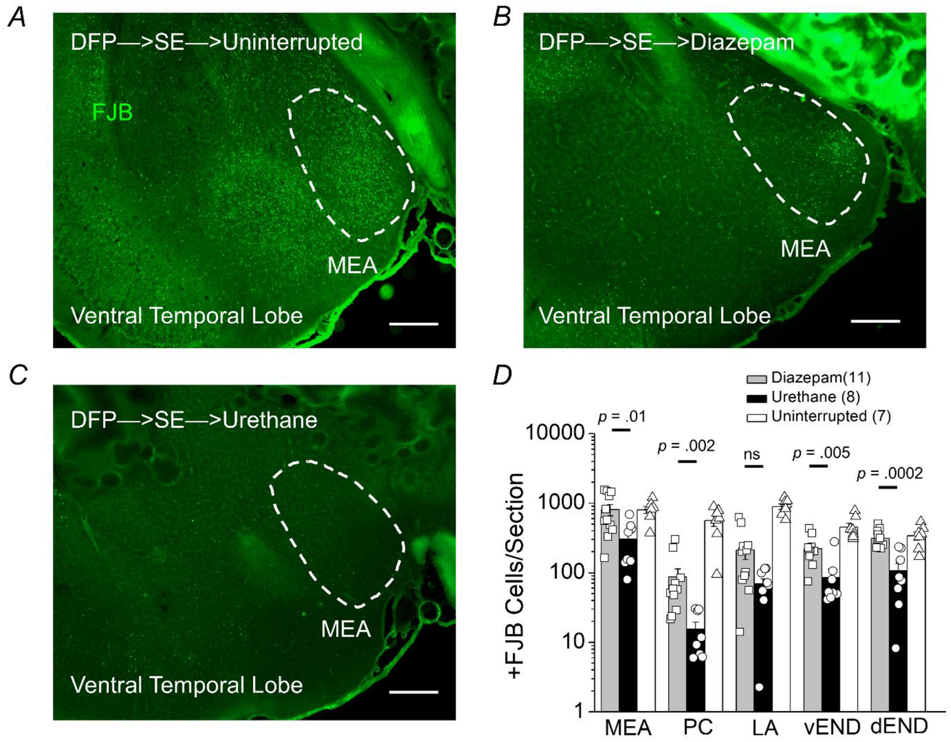
Representative images of FluoroJade B staining in sections (40 μm) of the ventral surface of the temporal lobe 24 hours after DFP-induced status epilepticus for rats that experienced uninterrupted SE (A), rats treated with diazepam after 1 h of SE (B), and rats injected with urethane after 1 h of SE (C). The images were taken at 25x (total magnification). The dashed circles outline the region of the medial amygdala (MEA). The images are representative of 5 sections from the ventral temporal lobe per rat. Scale bar = 500 μm. D, the average number of injured neurons per section 24 h after DFP-induced SE in five nuclei of the ventral temporal lobe (medial amygdala, MEA; piriform cortex, PC; lateral amygdala, LA; ventral endopiriform nucleus, vEND; dorsal endopiriform nucleus, dEND) of rats that experienced uninterrupted SE (white bar, n = 7 rats), rats treated with diazepam following 1 h of SE (gray bar, n = 11 rats) and rats injected with urethane after 1 h of SE (black bar, n = 8 rats). The bars show the mean and standard error of the mean. The symbols represent each individual rat within the group. The Mann-Whitney test was used to compare urethane to diazepam treated rats.