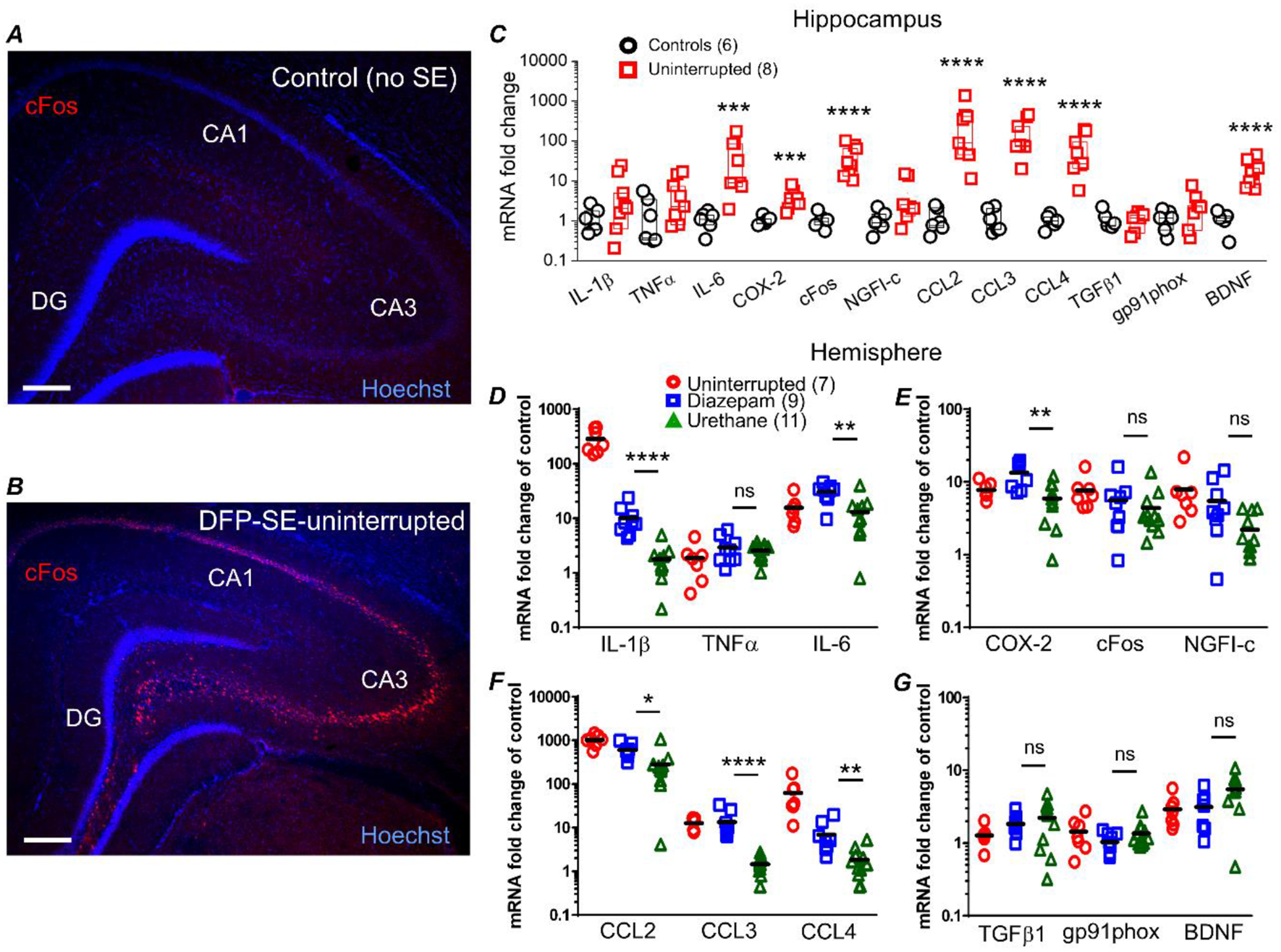
Figure 7. Urethane attenuates induction of brain inflammatory mediators 24 hours after SE.

Representative image of cFos staining (red) and Hoechst staining (blue) in a hippocampal section (40 μm) obtained from a non-seizure control rat (A) and a rat that experienced uninterrupted DFP-induced sacrificed at 24 h (B). The images were taken at 25x total magnification. The images shown are representative of five sections each from three rats per treatment. Scale bar = 350 μm. CA1, cornu ammonis 1; CA3, cornu ammonis 3; DG, dentate gyrus. qRT-PCR was used to quantify the change in abundance of mRNAs of inflammatory mediators, immediate early response factors and growth factors from rats 24 h after injection with water or DFP-induced SE. C, the mRNA fold change of interleukin 6 (IL-6), chemokine (C-C motif) ligand 2 (CCL2), chemokine (C-C motif) ligand 3 (CCL3), chemokine (C-C motif) ligand 4 (CCL4), cyclooxygenase 2 (COX-2), FBJ murine osteosarcoma viral oncogene homolog, (cFos) and brain derived neurotrophic factor (BDNF) were robustly upregulated in the hippocampus of rats that experienced uninterrupted DFP-induced SE. Although the fold changes are shown statistical analysis was carried out using the ΔΔCT values. ns = p >.05, *** = p < .001, **** = p < .0001, student’s t-test. The abundance of mRNA in the brain hemisphere of the 12 genes were separated into 4 panels with three genes shown per panel. IL-1β, interleukin 1 beta; TNFα, tumor necrosis factor alpha and IL-6 are shown in (D). COX-2, cFos and nerve growth factor-induced early-response gene (NGFI-c) are shown in (E). CCL2, CCL3 and CCL4 are shown in (F). Transforming growth factor beta-1 (TGFβ1), NADPH oxidase 2 (GP91phox) and BDNF are shown in (G). The difference in the level of upregulation of the hippocampus compared to the hemi-brain is likely due to contributions of other brain regions in the hemi-brains. The mRNA fold change of IL-1β, IL-6, CCL2, CCL3, CCL4 and COX-2 were significantly reduced in rats that received a single injection of urethane 1 h after SE compared to rats that received diazepam. Although the fold changes are shown statistical analysis was carried out using the ΔΔCT values. ns = p >.05, * = p < .05, ** = p < .01, **** = p < .0001, one-way ANOVA with posthoc Bonferroni (urethane vs. diazepam). The symbols represent each individual rat within the group. The black horizontal bar within the symbols represents the mean.
