Figure 4.

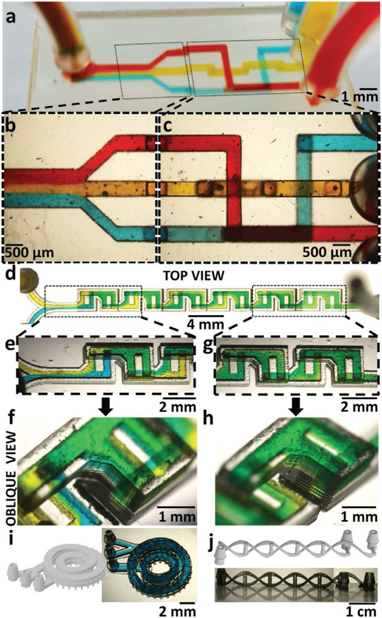
3D microfluidics. a) Oblique view of a 3D-printed 3-way microfluidic “3D-router” with red, yellow, and blue dye flowing through 500 µm square channels. b) Magnified top-view micrograph of the 3-way microfluidic router channels at the outlet area, showing the laminar flow of the three dyes as they merge in the outlet channel. c) Magnified top-view micrograph of the 3D-router showing the channels at the inlet area. The image shows channels crossing each other (blue and yellow, red and yellow) or overlaying each other (red and blue), without the dyes mixing. d) Top-view micrograph of a 3D-printed F-mixer (500 µm wide, 500 µm tall channels) with blue and yellow dyes flowing at 6 mL h−1. e) Top-view micrograph of the laterally inverted, stacked F-units next to the inlets. The fluid in the upper (second) F-unit appears green. In the subsequent lower F-unit, the fluid splits back into separate blue and yellow streams, confirming the vertical lamination of flow in the previous F-unit. f) Oblique-view micrograph of the F-units next to the outlet. The fluid in the last F-unit appears almost homogeneously green, confirming the effectiveness of this mixer design at mixing fluids. g) Top-view micrograph of the first F-unit showing the vertical lamination of blue and yellow dyes visible through the translucent side-walls of the device. h) Oblique-view micrograph of the last F-unit showing almost complete mixing of the blue and yellow dyes. i) (Left) 3D-CAD of a two-turn coil microchannel nested inside another two-turn coil microchannel. (Right) Oblique-view micrograph of the 3D-printed device filled with blue dye. The channels are 1 mm wide and 300 µm tall. j) (Top) 3D-CAD of two microchannels intertwined like a double-helix DNA, with the “nucleotide” side channels acting as points of fluidic contact between the two main channels. (Bottom) Side-view micrograph of the 3D-printed double-helix DNA microfluidic device. The main channels are 400 µm × 750 µm in cross section and the side channels are 500 µm in diameter.
