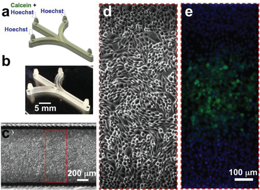
Figure 8.

Cell labeling in 3D-printed microchannels. a) CAD design of the microfluidic device for a simple cell labeling experiment. The device integrates three inlet and one outlet barb connectors. For focal labeling of the cells, Calcein Green AM (5 µm) and Hoechst 33342 (1 µm) were flowed through the middle inlet, while only Hoechst 33342 (1 µm) was introduced through the side inlets. b) Oblique-view photograph of the 3D-printed microfluidic device. c) Stitched phase-contrast micrograph of a confluent layer of Chinese hamster ovary (CHO-K1) cells growing in the device. d) Magnified phase-contrast micrograph of CHO-K1 cells in the channel. e) Magnified fluorescence micrograph of CHO-K1 cells in the channel focally labeled with Calcein Green AM. All the cells in the channel are also labeled with the nuclear dye, Hoechst 33342.
