Figure 1.

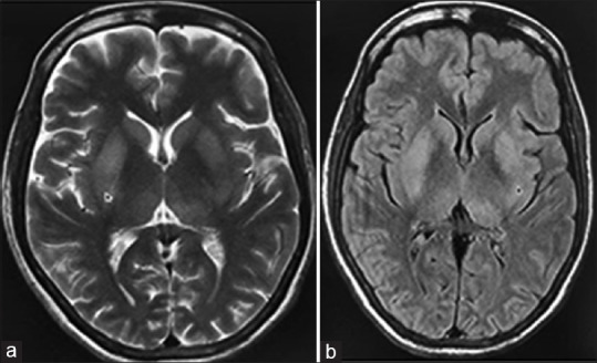
Magnetic resonance imaging of brain; (a) T2W image (b) FLAIR image showing mild hyperintensities in bilateral basal ganglia (arrows) without diffusion restriction or hemorrhages on the gradient (not shown)

Magnetic resonance imaging of brain; (a) T2W image (b) FLAIR image showing mild hyperintensities in bilateral basal ganglia (arrows) without diffusion restriction or hemorrhages on the gradient (not shown)