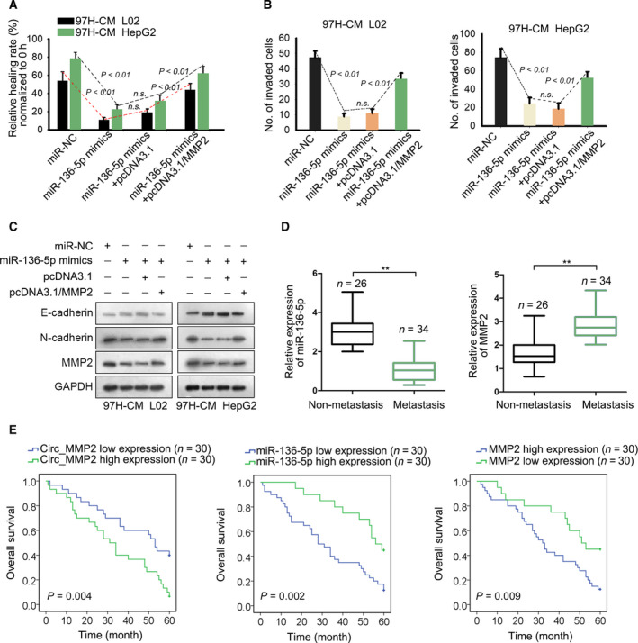
Figure 6.

MMP2 and miR‐136‐5p are crucial participants in HCC metastasis. (A, B) Cell migration and invasion were examined in 97H‐CM‐cultured L02 and HepG2 cell lines after transfecting with miR‐136‐5p mimics or cotransfected with pcDNA3.1/MMP2 (mean ± SD; n = 6; one‐way ANOVA). (C) The level of E‐cadherin, N‐cadherin, and MMP2 was examined in indicated cell lines. (D) Expression level of miR‐136‐5p or MMP2 in HCC tissues with or without metastasis (mean ± SD; n = 60; Student's t‐test). (E) Overall survival rate of HCC patients with high or low level of circ_MMP2, miR‐136‐5p, or MMP2 (Kaplan–Meier method followed by log‐rank test). P < 0.01 indicated data were statistically significant. n.s., no significance. **means P < 0.01.
