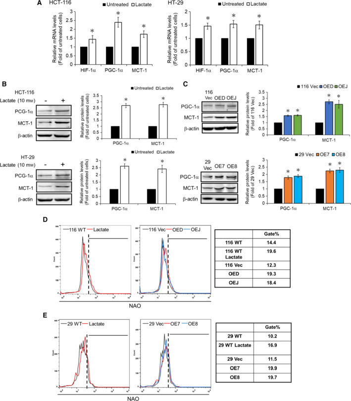
Figure 6.

Lactate is a critical mediator to promote the mitochondrial biogenesis in human colon cancer cells. (A) The wild‐type HCT‐119 (left) and HT‐29 (right) cells were treated without or with 10 mm lactate for 48 hr before the mRNA levels of HIF‐1α, PGC‐1α, and MCT‐1 were analyzed by RT‐qPCR. (B) Total lysates (30 µg) prepared from the wild‐type HCT‐119 (upper) and HT‐29 (lower) cells after similar treatments were subjected to immunoblot analysis using antibodies against PGC‐1α and MCT‐1 as probes, respectively. Beta‐actin signals were used as loading controls. Data are mean ± SD of three independent experiments. *P < 0.05 compared with those of the corresponding wild‐type cells by Student's t‐test. (C) Total lysates (30 µg) prepared from the 116 Vec, OED, and OEJ clones (upper) as well as the 29 Vec, OE7, and OE8 clones (lower) were subjected to immunoblot analysis using antibodies against PGC‐1α and MCT‐1 as probes, respectively. Beta‐actin signals were used as loading controls. Data are mean ± SD of three independent experiments. *P < 0.05 compared with those of the corresponding vector‐control clones by Student's t‐test. The mitochondrial mass of (D) the wild‐type HCT‐116 cells before and after treatment with lactate (left) as well as the 116 Vec, OED, and OEJ clones (middle), and (E) the wild‐type HT‐29 cells before and after treatment with lactate (left) as well as the 29 Vec, OE7, and OE8 clones (middle) were measured by flow cytometry after they were stained with 100 nm NAO. The gate population (right) was measured by flowjo V10 software.
