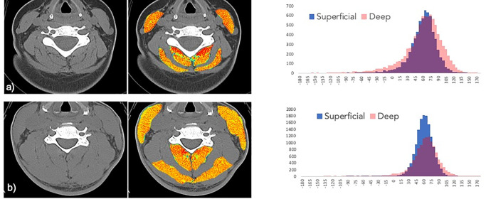
Fig 1. Radiation attenuation map of neck muscles at C5.
a) Subject 1 is a 32-year- old female with a body mass index of 25.8 kg m2 with poor recovery at 12-months post-MVC. The paraspinal muscles exhibit extensive visible fat within the fascia surrounding skeletal muscle making up 5.1% of total tissue area. Exclusive of the intermuscular fat, the mean overall radiation attenuation is 53.9 HU. b) Subject 2 is a 50-year-old male with a body mass index 27.5 kg m2 reporting full-recovery at 12-months post-MVC. There is much less visible regions of intermuscular fat infiltration (light blue) comprising 1.2% of total area, a value on the order of 4 fold lower than Subject 1. Exclusive of the macroscopic fat infiltration, the muscles show an overall mean attenuation of 59.0 HU. Corresponding histograms display the HU ranges and counts for the deep (pink) and superficial (blue) musculature (purple represents the HU overlap between the deep and superficial muscles).

