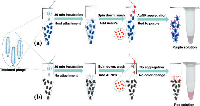
Figure 1.
Scheme for bacterial detection by phage AuNPs. The thiolated phages were added to a sample containing bacteria recognized by the phage (a) or not recognized by the phage (b). The cell–phage complexes (a) or nonhost cells (b) were separated from free phage by centrifugation. Resuspension of cell pellets containing thiolated phage-induced aggregation of AuNPs (a), producing a color change from red (AuNPs) to purple (aggregates of AuNPs), while nonhost cells do not cause aggregation of AuNPs and thus cause no color change (b).

