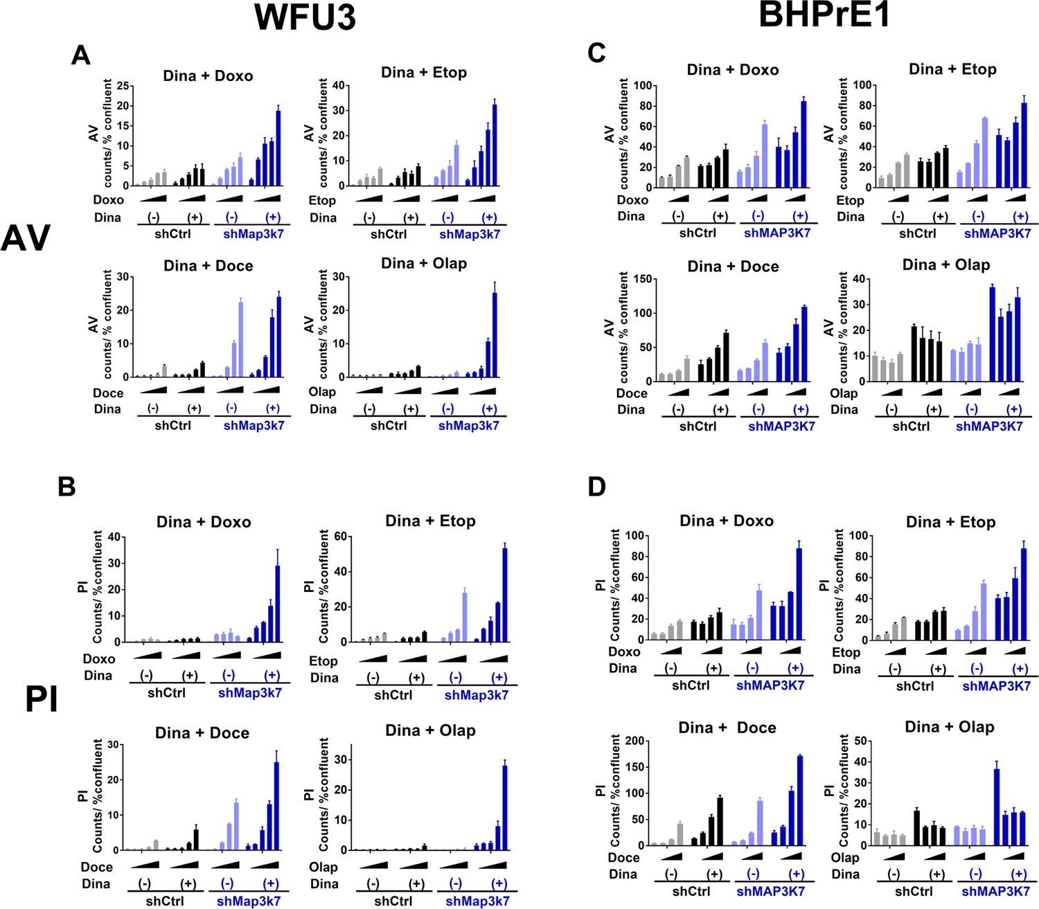
Figure 4: Dinaciclib augments effects of DNA damaging agents and olaparib in prostate cells with loss of MAP3K7.

A, B: Annexin V (A) and PI positive cells (B) in response to each DNA damaging agent or olaparib with or without dinaciclib in WFU3 shControl versus shMap3k7. Cells were treated with vehicle or each DNA damaging agent, doxorubicin 50–400nM, etoposide 0.63–5.0μM, docetaxel 1.25–10nM, or olaparib 1.25–10.0μM, with or without dinaciclib 60nM for 48hr. Annexin V and PI positive cells were assessed using IncuCyte Zoom and normalized to % confluence.
C, D: Annexin V (C) and PI positive cells (D) in BHPrE1 shControl versus shMAP3K7 in response to vehicle or each DNA damaging agent, doxorubicin 50–200nM, etoposide 1.0–4.0μM, docetaxel 0.25–1nM or olaparib 2.5–10.0μM with or without dinadicilib 4nM are shown. Cells were treated with these drugs for 48hr. Annexin V and PI positive cells were assessed using IncuCyte Zoom and normalized to % confluence.
Data were analyzed using two-way ANOVA and F ratio of column factor (each DNA damaging agent/ olaparib vs dinaciclib + each DNA damaging agent/olaparib) and p-value are also shown (Supplemental Table 12). Data are shown by mean and SE (n=3 per group). These results are representative of three independent experiments.
