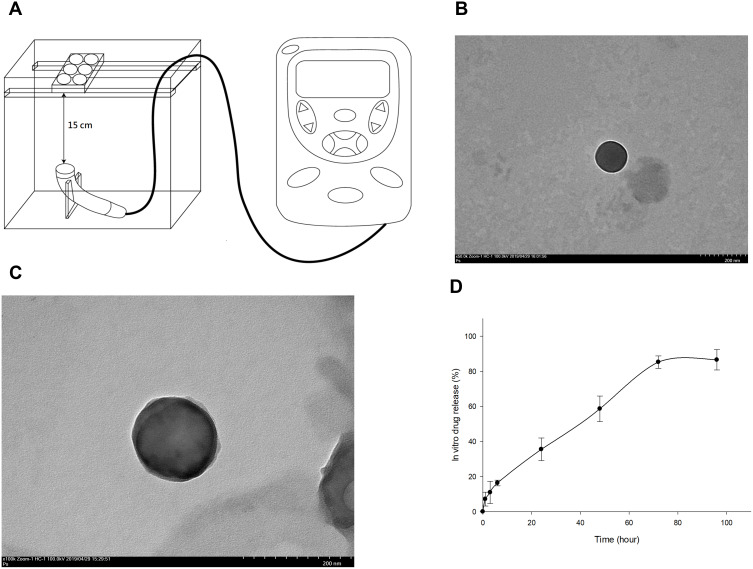
Figure 1.
(A) LIPUS was transmitted through a 15-cm water layer between the ultrasonic transducer and the bottom of culture plates. TEM image of prepared L-rapa showed an average of 135.17±28.3 nm at magnification 50,000× and 100,000× in (B) and (C), respectively (scale bar = 200 nm in both (B) and (C)). (D) In vitro release profile of rapamycin from rapamycin-loaded liposomes (N=3).

